. 2008 Nov 28;283(48):33026-35.
doi: 10.1074/jbc.M803646200. Epub 2008 Oct 6.

Receptor tyrosine phosphatase beta (RPTPbeta) activity and signaling are attenuated by glycosylation and subsequent cell surface galectin-1 binding

Affiliations

Receptor tyrosine phosphatase beta (RPTPbeta) activity and signaling are attenuated by glycosylation and subsequent cell surface galectin-1 binding

Karen L Abbott et al. J Biol Chem. .

Abstract

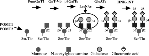
O-Mannosyl-linked glycosylation is abundant within the central nervous system, yet very few glycoproteins with this glycan modification have been identified. Congenital diseases with significant neurological defects arise from inactivating mutations found within the glycosyltransferases that act early in the O-mannosyl glycosylation pathway. The N-acetylglucosaminyltransferase known as GnT-Vb or -IX is highly expressed in brain and branches O-mannosyl-linked glycans. Our results using SH-SY5Y neuroblastoma cells indicate that GnT-Vb activity promotes the addition of the O-mannosyl-linked HNK-1 modification found on the developmentally regulated and neuron-specific receptor protein-tyrosine phosphatase beta (RPTPbeta). These changes in glycosylation accompany decreased cell-cell adhesion and increased rates of migration on laminin. In addition, we show that expression of GnT-Vb promotes its dimerization and inhibits RPTPbeta intrinsic phosphatase activity, resulting in higher levels of phosphorylated beta-catenin, suggesting a mechanism by which GnT-Vb glycosylation couples to changes in cell adhesion. GnT-Vb-mediated glycosylation of RPTPbeta promotes galectin-1 binding and RPTPbeta levels of retention on the cell surface. N-Acetyllactosamine, but not sucrose, treatment of cells results in decreased RPTP retention, showing that galectin-1 binding contributes to the increased retention after GnT-Vb expression. These results place GnT-Vb as a regulator of RPTPbeta signaling that influences cell-cell and cell-matrix interactions in the developing nervous system.


Figures

FIGURE 1.
FIGURE 1.
Proposed pathway for the production of O-mannosyl-linked HNK-1 epitope. POMT1/POMT2 catalyzes the transfer of mannose to a serine or threonine, followed by PomGnT1 adding GlcNAc in β1,2 linkage. GnT-Vb requires the PomGnT1 addition before it can add GlcNAc inβ1,6 linkage. After the addition of galactose residues by β4-galactosyltransferases (β4GalTs), the GlcATs (GlcATsS or GlcATsP) and the HNK-1 sulfotransferase (HNK1ST) form the HNK-1 epitope. LacNac the preferred substrate for a family of lectins known as galectins is circled.
FIGURE 2.
FIGURE 2.
GnT-Vb expression increases the production of O-mannosyl-linked HNK-1 epitope. A, WGA-precipitated proteins (Ppt) were analyzed by Western blot (WB) using the Cat-315 and CD57 antibodies. B, WGA immunoprecipitation reactions from GnT-Vb-expressing cell lysates were treated with the indicated enzymes before being analyzed by Western blot using the Cat-315 antibody.
FIGURE 3.
FIGURE 3.
GnT-Vb expression increases cell migration and reduces calcium-dependent cell-cell adhesion. A, confluent mock and GnT-Vb-expressing cells on laminin-coated chamber slides were wounded with a scratch, and subsequent migration was monitored for 4 and 6 h; bar 100 μm; arrows denotes wound boundary and direction of migration. Experiment shown is representative of three separate experiments. B, mock and GnT-Vb-expressing cells were removed from culture plates and separated into single-cell suspensions in calcium containing-media with constant agitation at 37 °C. Phase contrast microscopy after 30 min of a representative field; bar 50 μm. C, cumulative results from three separate experiments counted after 10 min of constant agitation at 37 °C. Error bars represent the mean percentage (±S.D.) of cell-cell adhesion from five randomly selected fields per experiment.
FIGURE 4.
FIGURE 4.
GnT-Vb glycosylates RPTPβ and inhibits intrinsic phosphatase activity in vitro and in vivo. A, upper box, total cell lysate (500 μg) from mock and GnT-Vb-expressing SH-SY5Y was immunoprecipitated (IP) using the anti-RPTPβ antibody, followed by Western blot (WB) analysis using the Cat-315 antibody. Lower box, anti-RPTPβ antibody Western blot analysis of the immunoprecipitations. B, tyrosine phosphatase activity assays using mock, GnT-Vb expressing, and GnT-Vb siRNA SH-SY5Y cells. RPTPβ was immunoprecipitated, and phosphatase activity was measured after 5 min using a tyrosine-phosphorylated peptide substrate (Promega). C, mock, GnT-Vb-expressing, and Gnt-Vb siRNA SH-SY5Y cells were serum-starved for 24 h before the addition of pleiotrophin at 50 ng/ml for 10 min. Lysates (500 μg) were immunoprecipitated with anti-β-catenin antibody, separated on 4–12% BisTris gels, and probed by Western blot for phosphotyrosine using the G410 antibody. Blots were re-probed for total β-catenin levels using anti-β-catenin antibody for loading normalization. Results shown are the average percentage increase in phospho-β-catenin levels following PTN treatment of GnT-Vb-expressing cells and mock-transfected cells from three separate experiments.
FIGURE 5.
FIGURE 5.
GnT-Vb glycosylation promotes RPTPβ cell-surface retention through a galectin-1 binding mechanism. A, RPTPβ cell-surface half-life was measured following cell surface biotinylation. Cells were re-cultured for 3 and 12 h before making cell lysates and pulling down biotinylated proteins by streptavidin magnetic bead precipitation, followed by detection of RPTPβ by Western blot using anti-RPTPβ antibody. B, RPTPβ dimerization was analyzed by chemical cross-linking using BS3 as described under “Experimental Procedures.” Total cell lysates (100 μg) were separated on a 3–8% Tris acetate gel before Western blot detection of RPTPβ. The results shown are representative of three separate experiments. C, densitometry analysis of dimerization experiments. Error bars represent the means (±S.D.) of the density of the dimer band normalized to monomer band density. D, galectin-1 cell-surface half-life was measured as described for A, except that anti-galectin-1 antibody was used following streptavidin magnetic bead precipitation. E, RPTPβ association with galectin-1 was assayed by protein cross-linking using BS3 as described under “Experimental Procedures” followed by immunoprecipitation (IP) and Western blotting using the indicated antibodies.
FIGURE 6.
FIGURE 6.
Inhibition of galectin-1 binding reduces cell-surface retention of RPTPβ. A, galectin-1 binding to the cell surface was measured by flow cytometry after the addition of biotinylated galectin-1 to suspended mock (shaded) or GnT-Vb-expressing (white) cells at 4 °C for 30 min in the presence of 10 mm sucrose (left) or 10 mm LacNAc (right). Bound biotinylated galectin-1 was detected using streptavidin-phycoerythrin. B, RPTPβ cell surface expression was monitored by flow cytometry after incubating mock (shaded) or GnT-Vb-expressing (white) cells for 2 h in the presence of 10 mm sucrose (left) or 10 mm LacNAc (right). Surface RPTPβ levels were detected on intact cells using an antibody to the extracellular region of RPTPβ, followed by detection using anti-IgM Alexa Fluor 633 secondary. Data presented have been subtracted for secondary-only fluorescence and are representative of two separate experiments.
FIGURE 7.
FIGURE 7.
Working model for the inhibition of RPTPβ activity and subsequent increases in β-catenin phosphorylation by GnT-Vb-mediated glycosylation and galectin-1 binding. A, in the absence of significant levels of O-mannosylated N-acetyllactosamine structures, which may include the HNK-1 epitope, RPTPβ is predominantly an active monomer. The active form of RPTPβ promotes the dephosphorylation (Y to Y) of tyrosine residues in β-catenin, causing more cell-cell adhesion and less cell motility. B, expression of GnT-Vb leads to more N-acetyllactosamine structures, including the HNK-1 epitope, that are bound by galectin-1. Galectin-1 binding stabilizes RPTPβ on the cell surface, promoting the dimerization of the receptor. RPTPβ dimerization then inhibits the phosphatase activity, leading to increased tyrosine phosphorylation (Y) of β-catenin that promotes the dissociation of the catenin-cadherin complex, resulting in decreased cell-cell adhesion and increased cell motility.

References

    1. Breen, K. C., Coughlan, C. M., and Hayes, F. D. (1998) Mol. Neurobiol. 16 163–220 - PubMed
    1. Schwarting, G. A., Jungalwala, F. B., Chou, D. K., Boyer, A. M., and Yamamoto, M. (1987) Dev. Biol. 120 65–76 - PubMed
    1. Bronner-Fraser, M. (1986) Dev. Biol. 115 44–55 - PubMed
    1. Abo, T., and Balch, C. M. (1981) J. Immunol. 127 1024–1029 - PubMed
    1. Lipinski, M., Braham, K., Caillaud, J. M., Carlu, C., and Tursz, T. (1983) J. Exp. Med. 158 1775–1780 - PMC - PubMed

Publication types

MeSH terms