. 2009 Jan;77(1):266-73.
doi: 10.1128/IAI.00402-08. Epub 2008 Oct 6.

Analysis of the specificity of Panton-Valentine leucocidin and gamma-hemolysin F component binding

Affiliations

Analysis of the specificity of Panton-Valentine leucocidin and gamma-hemolysin F component binding

Florent Meyer et al. Infect Immun. 2009 Jan.

Abstract

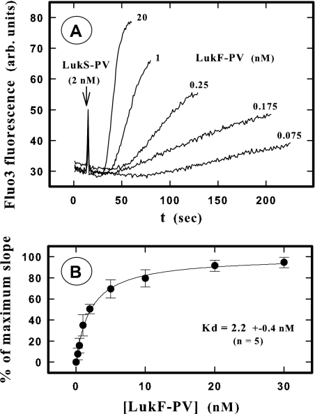
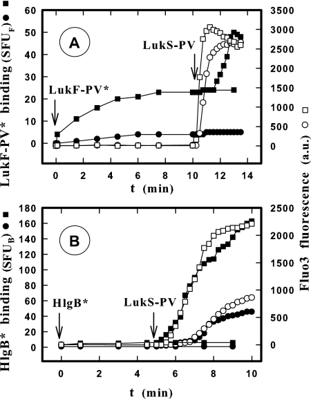
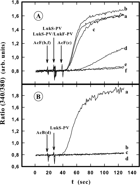
In this study, the binding of F components of the staphylococcal bicomponent leukotoxins Panton-Valentine leucocidin (LukF-PV) and gamma-hemolysin (HlgB) on polymorphonuclear neutrophils (PMNs), monocytes, and lymphocytes was determined using labeled mutants and flow cytometry. Leukotoxin activity was evaluated by measuring Ca(2+) entry or pore formation using spectrofluorometry or flow cytometry. Although HlgB had no affinity for cells in the absence of an S component, LukF-PV had high affinity for PMNs (dissociation constant [K(d)], 6.2 +/- 1.9 nM; n = 8), monocytes (K(d), 2.8 +/- 0.8 nM; n = 7), and lymphocytes (K(d), 1.2 +/- 0.2 nM; n = 7). Specific binding of HlgB was observed only after addition of LukS-PV on PMNs (K(d), 1.1 +/- 0.2 nM; n = 4) and monocytes (K(d), 0.84 +/- 0.31 nM; n = 4) or after addition of HlgC on PMNs, monocytes, and lymphocytes. Addition of LukS-PV or HlgC induced a second specific binding of LukF-PV on PMNs. HlgB and LukD competed only with LukF-PV molecules bound after addition of LukS-PV. LukF-PV and LukD competed with HlgB in the presence of LukS-PV on PMNs and monocytes. Use of antibodies and comparisons between binding and activity time courses showed that the LukF-PV molecules that bound to target cells before addition of LukS-PV were the only LukF-PV molecules responsible for Ca(2+) entry and pore formation. In contrast, the active HlgB molecules were the HlgB molecules bound after addition of LukS-PV. In conclusion, LukF-PV must be linked to LukS-PV and to a binding site of the membrane to have toxin activity.


Figures

FIG. 1.
FIG. 1.
Determination by flow cytometry of the binding of LukF-PV* and HlgB* labeled with fluorescein before and after addition of LukS-PV on human PMNs from the same donor. ▪, binding after 10 min of incubation for different concentrations of LukF-PV* (A) and HlgB* (B) with PMNs; •, additional binding 10 min after addition of 2 nM LukS-PV. Equilibrium constants calculated by nonlinear regression are shown in Tables 1 and 2. The slope of HlgB* binding obtained in the absence of LukS-PV is 6.05 ± 0.13 (mean ± standard deviation; n = 4). SFUF, standardized fluorescent units for LukF-PV*; SFUB, standardized fluorescent units for HlgB*.
FIG. 2.
FIG. 2.
Spectrofluorometric determination of the Kd of LukF-PV binding on PMNs by recording the time course of the increase in the intracellular Ca2+ level after addition of LukS-PV. (A) Fluorescence intensity for PMNs loaded with Fluo3 after 10 min of incubation with different LukF-PV concentrations and then addition of 2 nM LukS-PV. Only some of the concentrations used in one of five experiments are shown for clarity. arb. units, arbitrary units. (B) Plot of the higher slope values calculated for each LukF-PV concentration by nonlinear regression by the PTI software. The slopes were expressed as percentages of the maximal slope (100%) calculated for each experiment by nonlinear regression (single rectangular hyperbola plus nonspecific component) using the Sigma plot software (Systat Software Inc., San Jose, CA).
FIG. 3.
FIG. 3.
Flow cytometry determination of the competition between LukF-PV* and F components (LukF-PV, HlgB, and LukD) for binding in human PMNs (A), monocytes (B), and lymphocytes (C) in the presence and in the absence of LukS-PV. LukF-PV* at a concentration of 5 nM and different concentrations of F components were simultaneously added 10 min after 2 nM LukS-PV was added. The binding of 5 nM LukF-PV* was defined as 100%. The leukocyte concentration was 5 × 10⁴ cells/ml, and the incubation time was 15 min. The IC50s were as follows (means ± standard deviations): (A) 19.2 ± 2.2 nM for LukF-PV and 18.2 ± 4.2 nM for LukS-PV-LukF-PV (n = 6); (B) 14.1 ± 1.6 nM for LukF-PV (n = 4); (C) 7.1 ± 1.6 nM for LukF-PV (n = 4).
FIG. 4.
FIG. 4.
Flow cytometry determination of the competition between HlgB* and F components (HlgB, LukF-PV, and LukD) for binding in human PMNs (A) and monocytes (B) in the presence of LukS-PV. HlgB* at a concentration of 2 nM and different concentrations of F components were simultaneously added 10 min after 2 nM LukS-PV was added. The binding of 2 nM HlgB* was defined as 100%. The incubation time was 15 min. The IC50s were as follows (means ± standard deviations): (A) 2.3 ± 0.3 nM for HlgB (n = 5), 38.8 ± 1.6 nM for LukF-PV (n = 3), and 26.0 ± 1.6 nM for LukD (n = 4); (B) 1.5 ± 0.5 nM for HlgB (n = 3) and 9.5 ± 1.2 nM for LukF-PV (n = 3), and 20.4 ± 1.2 nM for LukD (n = 3).
FIG. 5.
FIG. 5.
Flow cytometry comparison of binding time courses (• and ▪) for LukF-PV* (A) and HlgB* (B) and increases in the intracellular Ca2+ level (○ and □) before and after addition of LukS-PV on PMNs. The LukF-PV* concentrations were 1 (○ and •) and 5 nM (□ and ▪), and the HlgB* concentrations were 0.3 (○ and •) and 1.2 nM (□ and ▪). LukS-PV (2 nM) was added 10 min after LukF-PV* was added (A) and 5 min after HlgB* was added (B), when binding was observed. The PMNs (5 × 10⁴ cells/ml) used for both determinations were from the same donor and were loaded with Fluo3. The LukF-PV and HlgB concentrations used were lower than and close to the apparent Kds. The experiment was carried out four times, and the same results were obtained each time. Therefore, the results of only one experiment are shown. SFUF, standardized fluorescent units for LukF-PV*; SFUB, standardized fluorescent units for HlgB*; a.u., arbitrary units.
FIG. 6.
FIG. 6.
Spectrofluorometric determination of anti-LukF-PV (A) and anti-HlgB (B) antibody influence on the increase in the intracellular Ca2+ level induced in the presence of LukF-PV and HlgB following addition of LukS-PV in Fluo3-containing human PMNs. (A) PMNs were incubated for 10 min with 5 nM LukF-PV before addition of 2 nM LukS-PV (lines a, b, c, d, and e), and LukF-PV and LukS-PV were also added simultaneously (line f). Anti-LukF-PV was added as follows: line a, no addition; line b, 10 s before addition of LukS-PV; line c, 10 s after addition of LukS-PV; line d, 5 min before addition of LukS-PV; line e, 10 s before addition of LukF-PV; line f, 10 s before addition of LukF-PV-LukS-PV. (B) PMNs were incubated for 10 min with 2 nM HlgB before addition of 2 nM LukS-PV (lines a, b, c, and d). Anti-HlgB was added as follows: line a, no addition; line b, 10 s before addition of LukS-PV; line c, 10 s before addition of HlgB; line d, 5 min before addition of LukS-PV. The experiment was done four times, and the same results were obtained each time. arb. units, arbitrary units.
FIG. 7.
FIG. 7.
Comparison by flow cytometry of LukF-PV* binding induced by LukS-PV and pore formation obtained under the same conditions. (A) Binding of LukF-PV* after addition of different concentrations of LukS-PV to PMNs after 10 min of incubation with 5 nM LukF-PV*. (B) Pore formation measured by ethidium fluorescence after addition of LukS-PV under the conditions described above for panel A and expressed as percentages of the maximal fluorescence intensity. The maximal fluorescence intensity (100%), corresponding to the maximal possible entry of ethidium into PMNs, was calculated by nonlinear regression of a sigmoidal model (SigmaPlot). The experiment was carried out three times, and the same results were obtained each time. The results of one experiment are shown.
FIG. 8.
FIG. 8.
Flow cytometry determination of the time course of the increase in the intracellular Ca2+ level and pore formation induced by LukF-PV in the presence of LukS-PV or HlgA in human monocytes and lymphocytes. The intracellular Ca2+ level was determined by Fluo3 fluorescence, and pore formation was evaluated using ethidium fluorescence with 5 × 10³ mixed monocytes and lymphocytes. Cells were incubated with 5 nM LukF-PV for 10 min before addition of 2 nM LukS-PV or 5 nM HlgA at time zero. arb. units, arbitrary units.
FIG. 9.
FIG. 9.
Schematic diagram of the association of PVL components and HlgB. (Panel I) LukS-PV (S) and LukF-PV (F) bind to their membrane receptors (RS and RF, respectively) and associate to open a Ca2+ channel (C) and form a pore (P) through the membrane (M). (Panel II) LukF-PV binds to the LukS-PV-LukS-PV membrane receptor complex, forming an inactive association. (Panel III) HlgB binds to the LukS-PV-LukS-PV membrane receptor complex to open a calcium channel and form a pore through the membrane.

References

    1. Baba Moussa, L., S. Werner, D. A. Colin, L. Mourey, J. D. Pédelacq, J. P. Samama, A. Sanni, H. Monteil, and G. Prévost. 1999. Discoupling the Ca2+-activation from the pore-forming function of the bi-component Panton-Valentine leucocidin in human PMNs. FEBS Lett. 461280-286. - PubMed
    1. Bocchini, C. E., K. G. Hulten, E. O. Mason, Jr., B. E. Gonzalez, W. A. Hammerman, and S. L. Kaplan. 2006. Panton-Valentine leukocidin genes are associated with enhanced inflammatory response and local disease in acute hematogenous Staphylococcus aureus ostomyelitis in children. Pediatrics 117433-440. - PubMed
    1. Boyle-Vavra, S., and R. S. Daum. 2007. Community-acquired methicillin-resistant Staphylococcus aureus: the role of Panton-Valentine leukocidin. Lab. Investig. 873-9. - PubMed
    1. Colin, D. A., I. Mazurier, S. Sire, and V. Finck-Barbançon. 1994. Interaction of the two components of leukocidin from Staphylococcus aureus with human polymorphonuclear leukocyte membranes: sequential binding and subsequent activation. Infect. Immun. 623184-3188. - PMC - PubMed
    1. Colin, D. A., and H. Monteil. 2003. Control of the oxidative burst of human neutrophils by staphylococcal leukotoxins. Infect. Immun. 713724-3729. - PMC - PubMed

Publication types

MeSH terms

LinkOut - more resources