Comparative Study
. 2008 Oct 27;205(11):2473-81.
doi: 10.1084/jem.20081241. Epub 2008 Oct 6.

Aquaporin-4-binding autoantibodies in patients with neuromyelitis optica impair glutamate transport by down-regulating EAAT2

Affiliations
Comparative Study

Aquaporin-4-binding autoantibodies in patients with neuromyelitis optica impair glutamate transport by down-regulating EAAT2

Shannon R Hinson et al. J Exp Med. .

Abstract

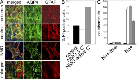
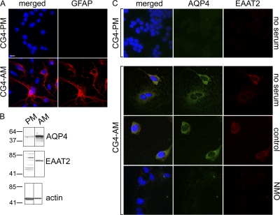
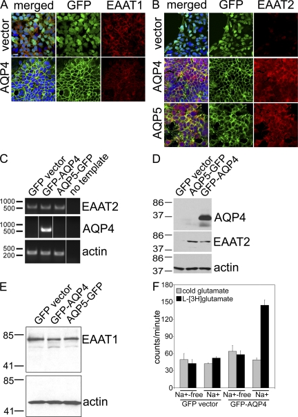
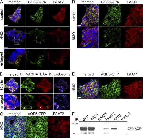
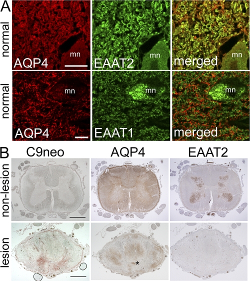
Neuromyelitis optica (NMO)-immunoglobulin G (IgG) is a clinically validated serum biomarker that distinguishes relapsing central nervous system (CNS) inflammatory demyelinating disorders related to NMO from multiple sclerosis. This autoantibody targets astrocytic aquaporin-4 (AQP4) water channels. Clinical, radiological, and immunopathological data suggest that NMO-IgG might be pathogenic. Characteristic CNS lesions exhibit selective depletion of AQP4, with and without associated myelin loss; focal vasculocentric deposits of IgG, IgM, and complement; prominent edema; and inflammation. The effect of NMO-IgG on astrocytes has not been studied. In this study, we demonstrate that exposure to NMO patient serum and active complement compromises the membrane integrity of CNS-derived astrocytes. Without complement, astrocytic membranes remain intact, but AQP4 is endocytosed with concomitant loss of Na(+)-dependent glutamate transport and loss of the excitatory amino acid transporter 2 (EAAT2) . Our data suggest that EAAT2 and AQP4 exist in astrocytic membranes as a macromolecular complex. Transport-competent EAAT2 protein is up-regulated in differentiating astrocyte progenitors and in nonneural cells expressing AQP4 transgenically. Marked reduction of EAAT2 in AQP4-deficient regions of NMO patient spinal cord lesions supports our immunocytochemical and immunoprecipitation data. Thus, binding of NMO-IgG to astrocytic AQP4 initiates several potentially neuropathogenic mechanisms: complement activation, AQP4 and EAAT2 down-regulation, and disruption of glutamate homeostasis.


Figures

Figure 1.
Figure 1.
In primary astrocytes, NMO-IgG induces internalization of AQP4, impairment of glutamate uptake, or complement activation. (A) Plasma membrane AQP4 (green) and cytoplasmic GFAP (red) after exposure to no human serum, control patient serum, or NMO patient serum. Serum containing NMO-IgG causes rapid translocation of surface AQP4 into cytoplasmic vesicles. Boxed area is enlarged in bottom images. Bars, 10 μm. (B) Quantitation of membrane permeability after exposure to control or NMO serum. Increase in permeability to propidium iodide (PI) greater than twofold by NMO serum required active complement (Δ C′ = inactivated complement). (C) Uptake of l-[3H]glutamate (±Na+-containing buffer) without human serum (white column) or in control (gray column) or NMO serum (crosshatched column). Excess unlabeled glutamate (black column) prevented l-[3H]glutamate uptake. NMO serum reduced l-[3H]glutamate uptake by 50%. The experiment shown in C was performed twice. All other experiments were done at least three times. The error bars represent the standard error of six and four replicates, respectively.
Figure 2.
Figure 2.
CG-4 glial cells in astrocytic differentiation medium up-regulate both AQP4 and EAAT2 expression; NMO serum down-regulates both. (A) CG-4 cells cultured in proliferation medium (CG4-PM) do not express cytoplasmic GFAP (red). After 7 d in astrocytic differentiation medium (CG4-AM), a subset of cells contains brilliant cytoplasmic GFAP. (B) The Western blot shows that undifferentiated CG4-PM (PM) cell lysates contain minimal EAAT2 or AQP4; both proteins are up-regulated in CG4-AM (AM). Markers indicate kilodalton reference standards. (C) AQP4 (green) and EAAT2 (red) are negligible in CG4-PM cells, but are up-regulated on the surface of CG4-AM cells; NMO serum, but not control patient serum, depletes both. DNA is blue (A and C). All experiments were performed a minimum of two times. Bars, 5 μm.
Figure 3.
Figure 3.
Expression of AQP proteins in HEK-293 cells induces EAAT2 protein expression. DNA is stained blue (A and B). (A) EAAT1 (red) is expressed constitutively, regardless of transfection. (B) EAAT2 (red) is localized in the plasma membrane of cells expressing AQP4 or AQP5 (green), but is undetectable in cells transfected with GFP-vector lacking an AQP cDNA. Bars, 10 μm. (C) For RT-PCR, all three cell lines express EAAT2 mRNA, regardless of AQP expression. Markers indicate basepair reference standards. (D) The Western blot shows that EAAT2 protein is up-regulated in cells transfected with AQP4 or AQP5, but not in GFP-vector–transfected cells. (E) EAAT1 protein and actin are expressed constitutively. Markers indicate kilodalton reference standards (D and E). (F) In glutamate transport experiments, GFP-AQP4 cells take up approximately threefold more l-[3H]glutamate than vector-transfected cells (note: Na+ dependence). All experiments were performed a minimum of two times. The error bars represent the standard error of six and four replicates, respectively.
Figure 4.
Figure 4.
NMO-IgG modulation of AQP4 from transfected HEK-293 membranes down-regulates EAAT2 expression. GFP-AQP4– or AQP5-GFP–transfected cells (green) were probed with anti-EAAT2 or –EAAT1-IgG (red) after exposure to control or NMO patient serum (37°C; 4 h). Nuclear DNA is blue. (A) NMO serum caused membrane loss of both AQP4 and EAAT2; control serum did not affect the distribution of either. Enlarged images show both EAAT2 and AQP4 internalized in cytoplasmic vesicles after exposure to NMO serum. (B) Staining with early endosome marker (EEA1, purple) after 10-min exposure to NMO serum shows colocalization of both GFP-AQP4 and EAAT2 (arrows) in endocytotic vesicles (white spots in merged image). (A and B) Boxed areas are enlarged in the bottom image. (C) NMO serum does not selectively affect EAAT2 in cells transfected with AQP5. EAAT1 is not affected by NMO or control serum in cells transfected with AQP4 (D) or AQP5 (E). (F) Western blot shows NMO-IgG or IgG specific for GFP, AQP4, or EAAT2, capture GFP-AQP4 protein; GFP-AQP4 does not coprecipitate with control patient IgG or EAAT1-specific IgG. Markers indicate kilodalton reference standards. All experiments were performed a minimum of two times. Bars: (A, C, D, and E) 10 μm; or (B) 20 μm.
Figure 5.
Figure 5.
Glutamate transporter expression in human tissue. (A) Normal spinal cord from a single subject. Duplicate sections were dual stained for AQP4 (red) and EAAT2 or EAAT1 (green), i.e., four sections. Merged images show AQP4 and EAAT2 colocalization (top, yellow), but no colocalization of AQP4 and EAAT1 (bottom); mn, motor neuron. (B) Spinal cord tissue from a single NMO-IgG–seropositive patient. Three sections of nonlesioned lumbar region (top) serve as staining control for lesioned cord (bottom). The lack of complement deposition (C9neo, brick red in lesioned cord, bottom) and high expression of AQP4 in both white and gray matter are typical of normal cord tissue; EAAT2 is highly enriched in gray matter. Prominent deposition of C9neo in gray matter of lesioned thoracic cord (bottom, same patient) corresponds to focal regions of AQP4 and EAAT2 loss in adjacent sections. AQP4 is partially retained in the white matter. Asterisk, central canal. Bars: (A) 20 μm; (B) 200 μm.

References

    1. Wingerchuk, D.M., V.A. Lennon, C.F. Lucchinetti, S.J. Pittock, and B.G. Weinshenker. 2007. The spectrum of neuromyelitis optica. Lancet Neurol. 6:805–815. - PubMed
    1. Lennon, V.A., T.J. Kryzer, S.J. Pittock, A.S. Verkman, and S.R. Hinson. 2005. IgG marker of optic-spinal multiple sclerosis binds to the aquaporin-4 water channel. J. Exp. Med. 202:473–477. - PMC - PubMed
    1. Nakamura, M., I. Miyazawa, K. Fujihara, I. Nakashima, T. Misu, S. Watanabe, T. Takahashi, and Y. Itoyama. 2008. Preferential spinal central gray matter involvement in neuromyelitis optica. An MRI study. J. Neurol. 255:163–170. - PubMed
    1. Roemer, S.F., J.E. Parisi, V.A. Lennon, E.E. Benarroch, H. Lassmann, W. Bruck, R.N. Mandler, B.G. Weinshenker, S.J. Pittock, D.M. Wingerchuk, and C.F. Lucchinetti. 2007. Pattern-specific loss of aquaporin-4 immunoreactivity distinguishes neuromyelitis optica from multiple sclerosis. Brain. 130:1194–1205. - PubMed
    1. Lucchinetti, C.F., R.N. Mandler, D. McGavern, W. Bruck, G. Gleich, R.M. Ransohoff, C. Trebst, B.G. Weinshenker, D. Wingerchuk, J.E. Parisi, and H. Lassmann. 2002. A role for humoral mechanisms in the pathogenesis of Devic's neuromyelitis optica. Brain. 125:1450–1461. - PMC - PubMed

Publication types

MeSH terms