Comparative Study
. 2008 Oct 27;205(11):2609-21.
doi: 10.1084/jem.20081370. Epub 2008 Oct 6.

TLR3 is an endogenous sensor of tissue necrosis during acute inflammatory events

Affiliations
Comparative Study

TLR3 is an endogenous sensor of tissue necrosis during acute inflammatory events

Karen A Cavassani et al. J Exp Med. .

Abstract

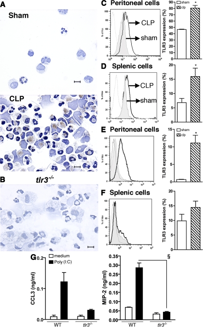
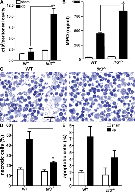
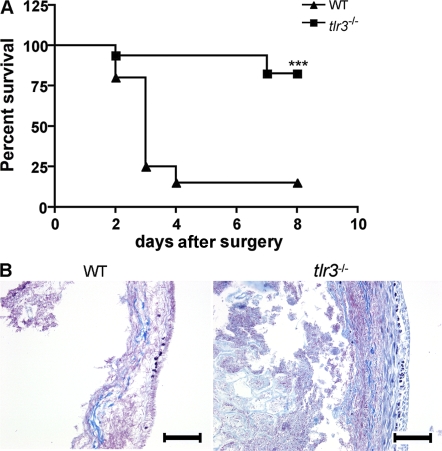
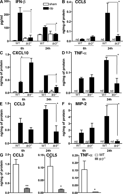
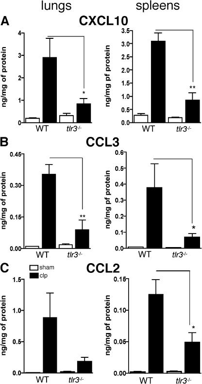
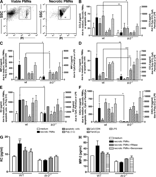
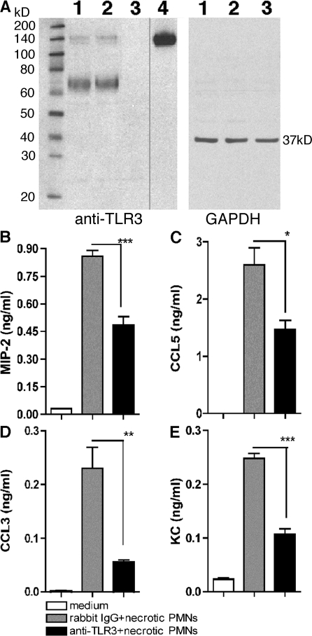
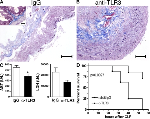
Ligands from dying cells are a source of Toll-like receptor (TLR) activating agents. Although TLR3 is known to respond to RNA from necrotic cells, the relative importance of this response in vivo during acute inflammatory processes has not been fully explored. We observed the involvement of TLR3 activation during experimental polymicrobial septic peritonitis and ischemic gut injury in the absence of an exogenous viral stimulus. In TLR3-deficient mice, increased chemokine/cytokine levels and neutrophil recruitment characterized the initial inflammatory responses in both injury models. However, the levels of inflammatory chemokines and tumor necrosis factor alpha quickly returned to baseline in tlr3(-/-) mice, and these mice were protected from the lethal effects of sustained inflammation. Macrophages from tlr3(-/-) mice responded normally to other TLR ligands but did not respond to RNA from necrotic neutrophils. Importantly, an immunoneutralizing antibody directed against TLR3 attenuated the generation of inflammatory chemokines evoked by byproducts from necrotic neutrophils cultured with wild-type macrophages. In vivo, anti-TLR3 antibody attenuated the tissue injury associated with gut ischemia and significantly decreased sepsis-induced mortality. Collectively, these data show that TLR3 is a regulator of the amplification of immune response and serves an endogenous sensor of necrosis, independent of viral activation.


Figures

Figure 1.
Figure 1.
CLP surgery induced local and systemic expression of TLR3. (A) Representative immunochemistry analysis of TLR3 expression in peritoneal cells from sham and CLP 6 h after surgery. TLR3 expression is observed in brown. Bars, 20 μm. (B) Immunochemistry analysis of TLR3 expression in thioglycollate-elicited macrophages of tlr3−/− mice. Bar, 20 μm. (C and D) Histograms exhibit TLR3 expression in permeabilized peritoneal (C) and splenic (D) CD11b+ cells. (E and F) Nonpermeabilized peritoneal (E) and splenic (F) CD11b+ cells (gray line, isotype control antibody; shaded, sham mice; black line, CLP mice). Data are means ± SEM from three independent experiments. (G) Immunoreactive CCL3 and MIP-2 supernatant levels from thioglycollate-elicited WT and tlr3−/− peritoneal neutrophils exposed to poly(I:C). *, P < 0.05 compared with sham mice; §, P < 0.05 compared with WT mice.
Figure 2.
Figure 2.
Increased peritoneal neutrophil influx and a marked reduction in necrotic peritoneal cells during septic peritonitis in tlr3−/− mice. (A) WT and tlr3−/− peritoneal cells at 24 h after sham and CLP surgeries. (B) MPO levels in peritoneal lavage samples at 24 h after either sham or CLP surgery. Data are means ± SEM from two separate experiments. (C) Toluidine blue staining of peritoneal cells from WT and tlr3−/− mice at 24 h after CLP surgery. Bars, 50 μm. (D and E) Apoptotic and necrotic cells were identified by annexin V+/PI and annexin V+/PI+ staining, respectively. The data represent means ± SEM (n = 5 mice per condition). *, P < 0.05; and **, P < 0.01 compared with C57BL/6 mice after CLP surgery.
Figure 3.
Figure 3.
tlr3−/− mice are resistant to lethality after CLP surgery and exhibit less tissue damage after gut ischemia. (A) Septic peritonitis was induced by CLP in WT (n = 20) and tlr3−/− (n = 17) mice. Mortality was monitored every 24 h for 8 d. ***, P < 0.0001 compared with the WT group. (B) Representative Masson's trichrome staining of cecum at 24 h after gut ischemia induced by total cecal ligation (n = 6). Bars, 100 μm.
Figure 4.
Figure 4.
Acute but not sustained peritoneal inflammation in tlr3−/− mice after CLP or gut ischemia. (A–F) At 6 and 24 h after CLP surgery, WT and tlr3−/− peritoneal cytokine and chemokine concentrations were measured by ELISA. (G) WT and tlr3−/− peritoneal cytokine and chemokine concentrations were measured by ELISA. The data represent means ± SEM obtained from three independent experiments for CLP and two independent experiments for gut ischemia. *, P < 0.05 compared with WT groups. nd, not detected.
Figure 5.
Figure 5.
TLR3 amplified systemic inflammatory responses and secondary tissue injury after septic peritonitis. (A–C) At 24 h after sham and CLP surgeries, chemokines were measured in lung and spleen homogenates using ELISA. Data are means ± SEM from two independent experiments (n = 4–5 mice per experiment). *, P < 0.05; and **, P < 0.01 compared with WT groups.
Figure 6.
Figure 6.
TLR3 was required for chemokine generation by peritoneal macrophages after co-culture with necrotic but not apoptotic cells. (A) The necrotic cells were identified by analyzing the side scatter (SSC) and PI+ staining. (B–F) Chemokine concentrations in the supernatants of WT or tlr3−/− peritoneal macrophages co-cultured with necrotic neutrophils, apoptotic splenocytes, poly(I:C), CpG-ODN, Pam3Cys, or LPS and quantified using ELISA and/or Bio-Plex. The data are means ± SEM from three to four combined experiments. *, P < 0.05; **, P < 0.01; and ***, P < 0.001 compared with tlr3−/− mice. (G and H) KC (G) and MIP-2 (H) protein levels in 96-well tissue culture plates containing WT or tlr3−/− macrophages and one of untreated, RNase-, or Benzonase-treated necrotic PMNs. **, P < 0.01 when necrotic PMNs were compared with medium; #, P < 0.05 when enzyme-treated necrotic PMNs were compared with nontreated necrotic PMNs.
Figure 7.
Figure 7.
TLR3 was required for chemokine generation by WT peritoneal macrophages after co-culture with necrotic neutrophils. (A) The specificity of polyclonal rabbit anti-TLR3 was determined by Western blot analysis of TLR3 expression in the following samples: lane 1, WT macrophages; lane 2, tlr7−/− macrophages; lane 3, tlr3−/− macrophages; and lane 4, recombinant TLR3. The black line indicates that intervening lanes have been spliced out. (B-E) Chemokine concentrations in the supernatants of WT peritoneal macrophages co-cultured with necrotic neutrophils with IgG or anti-TLR3 polyclonal antibody were quantified using Bio-Plex. The data are means ± SEM of triplicate wells and are representative of three independent experiments. *, P < 0.05; **, P < 0.01; and ***, P < 0.001 compared with IgG group.
Figure 8.
Figure 8.
Anti-TLR3 antibody markedly reduced cecal damage induced by gut ischemia and enhanced survival after the induction of severe sepsis. (A and B) Representative histological sections from WT mice that received either IgG (A) or anti-TLR3 (B) antibody during gut ischemia. The inset in A is a representative histological cross section of cecum from a naive WT mouse. Bars, 100 μm. (C) At 24 h after surgery, serum from WT mice subjected to cecal ischemia and either IgG or anti-TLR3 antibody treatments (n = 4 mice per group) were analyzed for AST and LDH. The data are means ± SEM. *, P < 0.05 compared with the IgG group. (D) WT mice received 3 mg of anti-TLR3 antibody or isotype control at 6 and 24 h after CLP surgery, and the survival rates were monitored for 54 h (n = 7 for the anti-TLR3 antibody group; n = 8 for the IgG group).

References

    1. Pinsky, M.R. 2004. Dysregulation of the immune response in severe sepsis. Am. J. Med. Sci. 328:220–229. - PubMed
    1. Cohen, J. 2002. The immunopathogenesis of sepsis. Nature. 420:885–891. - PubMed
    1. Matzinger, P. 1994. Tolerance, danger, and the extended family. Annu. Rev. Immunol. 12:991–1045. - PubMed
    1. Gallucci, S., M. Lolkema, and P. Matzinger. 1999. Natural adjuvants: endogenous activators of dendritic cells. Nat. Med. 5:1249–1255. - PubMed
    1. Sauter, B., M.L. Albert, L. Francisco, M. Larsson, S. Somersan, and N. Bhardwaj. 2000. Consequences of cell death: exposure to necrotic tumor cells, but not primary tissue cells or apoptotic cells, induces the maturation of immunostimulatory dendritic cells. J. Exp. Med. 191:423–434. - PMC - PubMed

Publication types