. 2008 Oct 6;183(1):19-28.
doi: 10.1083/jcb.200803133.

The keratin-binding protein Albatross regulates polarization of epithelial cells

Affiliations

The keratin-binding protein Albatross regulates polarization of epithelial cells

Masahiko Sugimoto et al. J Cell Biol. .

Abstract

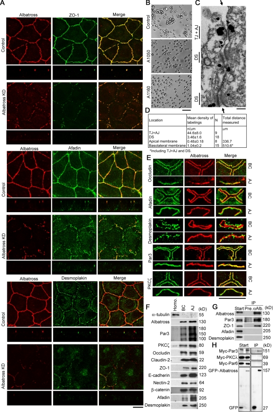
The keratin intermediate filament network is abundant in epithelial cells, but its function in the establishment and maintenance of cell polarity is unclear. Here, we show that Albatross complexes with Par3 to regulate formation of the apical junctional complex (AJC) and maintain lateral membrane identity. In nonpolarized epithelial cells, Albatross localizes with keratin filaments, whereas in polarized epithelial cells, Albatross is primarily localized in the vicinity of the AJC. Knockdown of Albatross in polarized cells causes a disappearance of key components of the AJC at cell-cell borders and keratin filament reorganization. Lateral proteins E-cadherin and desmoglein 2 were mislocalized even on the apical side. Although Albatross promotes localization of Par3 to the AJC, Par3 and ezrin are still retained at the apical surface in Albatross knockdown cells, which retain intact microvilli. Analysis of keratin-deficient epithelial cells revealed that keratins are required to stabilize the Albatross protein, thus promoting the formation of AJC. We propose that keratins and the keratin-binding protein Albatross are important for epithelial cell polarization.


Figures

Figure 1.
Figure 1.
Albatross is a keratin-binding protein. (A) Immunoblotting with affinity-purified rabbit anti-Albatross antibodies. Specific recognition of a polypeptide with a relative molecular mass of ∼130 kD, the putative size of Albatross, in HeLa and Caco-2 epithelial cells (middle). Preincubation of the antibody with the antigen selectively inhibited the immunoreactivity (right). CBB, a loading control (left). (B) Lysates prepared from the indicated cells were subjected to immunoblot analysis for Albatross. (C) Anti-Albatross antibody immunoprecipitants were subsequently immunoblotted with the indicated antibodies. Left and right lanes show immunoprecipitates with control rabbit IgG and anti-Albatross antibodies, respectively. With the indicated epithelial cells, Albatross, keratin 8, and keratin 18 immunoprecipitated as a complex. (D) Double staining of keratin 18 and Albatross. Albatross is discontinuously associated with the K8/18 filament network in the indicated epithelial cells. Mag., hyper-magnification of regions indicated by arrowheads. (E) Immunoblotting for keratin 8 and 18, and the indicated junctional and cell-polarity proteins in control and Albatross knockdown A549 (A1050 and A1160) cells. Albatross knockdown cells were produced successfully in which the expression level of keratin 8 and 18, the indicated AJC-specific transmembrane and undercoat proteins, lateral proteins, and apical proteins were not reduced. α-tubulin was included as a loading control. (F) Double staining for Albatross and keratin 18 in control and Albatross knockdown A549 (Albatross KD) cells. In control cells, Albatross is localized at the apical side of cell–cell border, and keratin filaments form apical bundles, whereas in Albatross KD cells, keratin filaments were apparently dissociated from cell–cell borders (arrowheads). Top and bottom columns illustrate projections of x-y planes and z sections, respectively. Bars, 10 μm.
Figure 2.
Figure 2.
Impaired AJC formation in Albatross knockdown cells. (A) Double staining for Albatross (red) and the undercoat proteins (green) for each AJC component: TJ, ZO-1; AJ, afadin; DS, desmoplakin. Top and bottom columns show projections of x-y planes and z sections, respectively. Albatross knockdown A549 (Albatross KD) cells lack accumulation of these proteins at the cell–cell borders except in regions where residual Albatross is present. (B) Cell–cell adhesive properties evaluated by a cell aggregation assay. In the differential interference contrast images, control cells show cell aggregation. With Albatross knockdown A549 (A1050 and A1160) cells, the aggregated cell population is reduced and free cells are increased. The percentages of single cells in total cells (mean ± SD) are: control, 36.1 ± 3.9; A1050, 52.4 ± 2.8; A1160 cells, 59.4 ± 10.2. n = 4 and P < 0.01. (C) Immunoelectron microscopy of A549 cells with anti-Albatross antibodies. Note that the cytoplasm in the vicinity of AJCs is labeled. TJ, AJ, and DS are indicated. Arrows indicate cell–cell contacts. (D) Quantitative data from C. (E) BC fraction and AJ fraction were immunostained for Albatross with the indicated AJC proteins, PKCζ or Par3. Note that Albatross is well colocalized with them. (F) Immunoblotting of fractions derived from mouse liver: homogenates (left), BC (middle), and AJ (right). Not only Albatross but also Par3 is enriched in line with the concentrations of the indicated AJC components. (G) Immunoprecipitation of A549 cells with anti-Albatross antibodies. Start and IP indicate starting lysates and immunoprecipitates with preimmune (Pre.) and anti-Albatross (αAlb.) antibodies, respectively. Note the Par3 precipitation with Albatross. Among AJC components, ZO-1 also coprecipitated. (H) Immunoprecipitation analysis with tagged Albatross and Par3. Start and IP indicate starting lysates and immunoprecipitates with anti-GFP antibodies, respectively. Left lanes show results for negative controls expressing GFP alone. Par3 was the most precipitated with GFP-Albatross among coexpressed myc-Par3, -Par6, and -PKCλ. Bars: (A) 10 μm; (B) 100 μm; (C) 0.1 μm; (E, BC) 13 μm; (E, AJ) 10 μm.
Figure 3.
Figure 3.
Albatross knockdown affects the localization of Par3 at AJCs and lateral proteins without affecting apical components. (A, B, and D–F) Double staining for Albatross and the indicated proteins. The top two rows indicate projections of x-y planes, and the bottom rows are for merged images of z sections. (A) Compared with control cells, Par3 is reduced in AJCs of Albatross knockdown A549 (Albatross KD) cells, though Par3 in the apical domains remains. (B) Ezrin is still localized in the apical domains of Albatross KD cells. (C) Scanning electron micrograph showing that microvilli on the apical domains are not impaired in Albatross KD (A1050 and A1160) cells. (D) In control cells, E-cadherin is localized on lateral domains and in AJCs. In Albatross KD cells, it is no longer accumulated on AJCs and lateral domains, and is mislocalized even at apical membranes. (E) Desmoglein 2 demonstrates similar alteration. (F) In control cells, cortical actin bundles are organized along the cell–cell border. In Albatross KD cells, they are not organized. Bars: (A, B, and D–F) 10 μm; (C) 1 μm.
Figure 4.
Figure 4.
Albatross–Par3 complex functions. (A) Rescue experiments. Double staining for Albatross and GFP, AJC components, or Par3 was performed with Albatross knockdown A549 cells in which GFP-Albatross or GFP (negative control) were introduced. Note that the GFP-Albatross–introduced cells show staining of GFP at cell–cell borders and recovery of the localization of Albatross, AJC components, and Par3. (B) Immunoblotting for the indicated junctional and cell-polarity proteins in control and Par3 knockdown A549 (A1555 and A1556) cells. Par3 knockdown cells have normal expression levels of the indicated AJC-specific proteins, lateral proteins, and apical proteins. GAPDH is included as a loading control. (C–E) Double staining for Par3 and the indicated proteins in Par3 knockdown A549 (Par3 KD) cells. The top two rows show projections of x-y planes and the bottom rows show merged images of z sections. (C) Compared with control cells, Albatross is reduced in AJCs of Par3 KD cells. (D) As in Albatross knockdown cells, desmoglein 2 no longer accumulated on AJCs and lateral domains, and was mislocalized even on the apical membranes in Par3 KD cells. (E) Par3 KD cells show impairment of apical localization of ezrin, in contrast to Albatross knockdown cells. Bars, 10 μm.
Figure 5.
Figure 5.
Functions of keratins and Albatross–Par3 complexes. (A–C) The amounts of Albatross protein and mRNA were analyzed in both keratin 8 and keratin 18 (K8/18)-introduced SW13 cells. As a control, an empty vector was transfected. As loading controls, α-tubulin and GAPDH were used. Two independent experiments were performed. (A) Immunoblotting. In transiently K8/18-introduced SW13 cells, the amount of Albatross protein is elevated, along with the amount of keratin 18. (B) With stable lines, the same results were obtained. (C) RT-PCR. In K8/18-introduced SW13 cells, the mRNA level of K18 is elevated, but not that of Albatross. β-actin is included as an internal control. (D) Double staining for K8/18 and the indicated proteins: Albatross, AJC components of ZO-1 and afadin, and Par3. (top) In control cells, K8/18 is absent and only limited amounts of Albatross are apparent at cell–cell junctions. In stably K8/18-introduced SW13 cells, Albatross is well localized in cell–cell junctions compared with control cells. (middle and bottom) ZO-1, afadin, and Par3 similarly accumulated at the cell–cell borders in stably K8/18-introduced SW13 cells. (E) Immunostaining of stably K8/18-introduced SW13 cells transfected with control or Albatross siRNA. Note that ZO-1, afadin, and Par3 are reduced at cell–cell borders with knockdown of Albatross. (F) A model for the regulation of AJC and lateral domains with the Albatross–Par3 complex and keratins. Albatross–Par3 complexes regulate the formation of AJC and maintain lateral membrane identity. However, Par3 without Albatross regulates apical structures. Keratins stabilize Albatross, promoting the formation of AJC. Knockdown effects are also indicated. Bars, 10 μm.

Similar articles

Cited by

References

    1. Bornslaeger, E.A., C.M. Corcoran, T.S. Stappenbeck, and K.J. Green. 1996. Breaking the connection: displacement of the desmosomal plaque protein desmoplakin from cell-cell interfaces disrupts anchorage of intermediate filament bundles and alters intercellular junction assembly. J. Cell Biol. 134:985–1001. - PMC - PubMed
    1. Caulin, C., C.F. Ware, T.M. Magin, and R.G. Oshima. 2000. Keratin-dependent, epithelial resistance to tumor necrosis factor-induced apoptosis. J. Cell Biol. 149:17–22. - PMC - PubMed
    1. Chen, X., and I.G. Macara. 2005. Par-3 controls tight junction assembly through the Rac exchange factor Tiam1. Nat. Cell Biol. 7:262–269. - PubMed
    1. Coulombe, P.A., and P. Wong. 2004. Cytoplasmic intermediate filaments revealed as dynamic and multipurpose scaffolds. Nat. Cell Biol. 6:699–706. - PubMed
    1. Fuchs, E., and D.W. Cleveland. 1998. A structural scaffolding of intermediate filaments in health and disease. Science. 279:514–519. - PubMed

Publication types

MeSH terms

Substances

Associated data