. 2008 Oct 6;183(1):129-41.
doi: 10.1083/jcb.200806104.

Notch activates cell cycle reentry and progression in quiescent cardiomyocytes

Affiliations

Notch activates cell cycle reentry and progression in quiescent cardiomyocytes

Víctor M Campa et al. J Cell Biol. .

Abstract

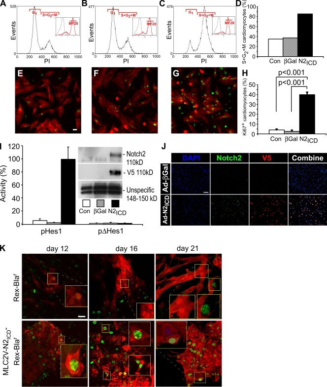
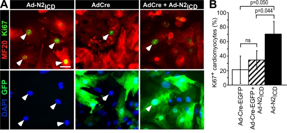
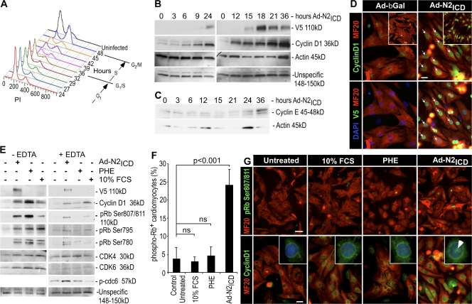
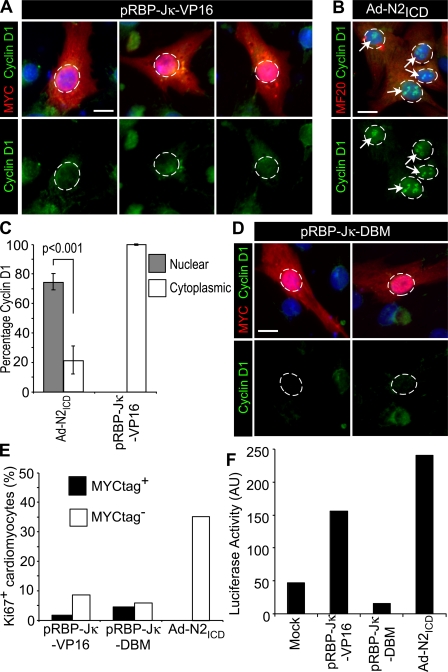
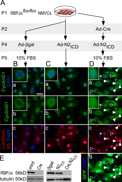
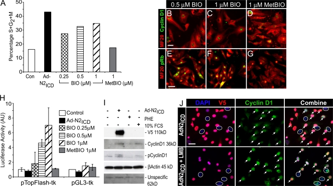
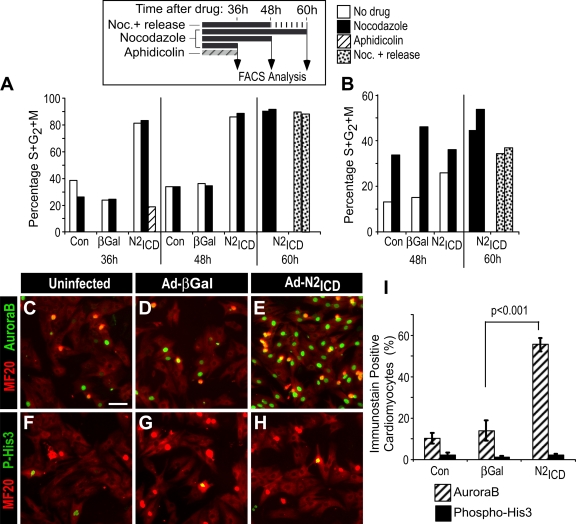
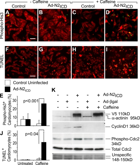
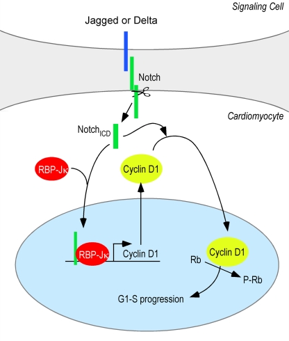
The inability of heart muscle to regenerate by replication of existing cardiomyocytes has engendered considerable interest in identifying developmental or other stimuli capable of sustaining the proliferative capacity of immature cardiomyocytes or stimulating division of postmitotic cardiomyocytes. Here, we demonstrate that reactivation of Notch signaling causes embryonic stem cell-derived and neonatal ventricular cardiomyocytes to enter the cell cycle. The proliferative response of neonatal ventricular cardiomyocytes declines as they mature, such that late activation of Notch triggers the DNA damage checkpoint and G2/M interphase arrest. Notch induces recombination signal-binding protein 1 for Jkappa (RBP-Jkappa)-dependent expression of cyclin D1 but, unlike other inducers, also shifts its subcellular distribution from the cytosol to the nucleus. Nuclear localization of cyclin D1 is independent of RBP-Jkappa. Thus, the influence of Notch on nucleocytoplasmic localization of cyclin D1 is an unanticipated property of the Notch intracellular domain that is likely to regulate the cell cycle in multiple contexts, including tumorigenesis as well as cardiogenesis.


Figures

Figure 1.
Figure 1.
Activated Notch induces cell cycle reentry in quiescent cardiomyocytes. (A–D) NRVCs were either uninfected (A and E) or infected with Ad-βGal (B and F) or Ad-N2ICD (C and G). 48 h after infection, cells were stained with mouse anti-MF20 (green, Alexa 488) and propidium iodide (PI; A–C). The number of MF20+ cells (green, Alexa 488) was >90% in all cases. DNA content was analyzed for the MF20+ population (insets) and the percentage in S/G2/M phase for each condition was calculated (D). The examples shown are representative of more than five experiments with similar outcomes. (E–H) Examples of NRVCs as in A–D stained with MF20 (red, Alexa 594) and Ki67 (green, Alexa 488; E–G) 48 h after infection, and the percentage of Ki67+ cells within the MF20+ population was measured (H). The examples shown are representative of more than five experiments with similar outcomes. (I) 12 h after infection, NRVCs were transfected with 0.6 μg of the pHes1-Luc or pΔHes1-Luc plasmids and 0.2 μg pGL3-Renilla-Luc. Luciferase activities and protein expression of Notch2 and V5 epitope (inset) were determined 12 h later. Firefly activity was normalized using Renilla luciferase activity. (J) Examples of NRVCs as in A–D stained with V5 (red, Alexa 594), Notch2, (green, Alexa 488), and DAPI (blue) 48 h after infection. (K) Confocal images showing the persistence of Ki67+ (green, Alexa 488) and MF20+ (red, Alexa 594) cardiomyocytes in differentiating mESCs expressing N2ICD from the MLC2V promoter compared with typically quiescent cardiomyocytes in cultures harboring only the REX promoter–Blar gene. Error bars indicate ±SD across experimental replicates. Bars: (E–G and K) 10 μm; and (J) 50 μm.
Figure 2.
Figure 2.
Cell cycle reentry requires RBP-Jκ. (A) Examples of RBP-JκFlox/Flox NMVCs infected with Ad-Cre-EGFP to remove RBP-Jκ, then reinfected 12 h later with Ad-N2ICD, as indicated, cultured 48 h, and stained with MF20 (red, Alexa 594), Ki67 (far red, Cy5, shown as green), and DAPI (blue). Arrows indicate Ki67+ cells. Bar, 10 μm. (B) The percentage of Ki67+ cardiomyocytes (MF20+) in each condition. Only Cre-expressing cells (GFP+, green) were counted in cultures infected with Ad-Cre-EGFP. N2ICD induced KI67 (P = 0.05) but the N2ICD effect following Cre was not significantly different from that of Cre alone (ns). Error bars indicate ±SD across experimental replicates.
Figure 3.
Figure 3.
Notch induces expression and nuclear localization of cyclin D1 and phosphorylation of Rb before cell cycle reentry. (A) The DNA content of the cardiac cells analyzed by flow cytometry (as in Fig. 1) at the indicated times after infection. One of two experiments with similar outcomes is shown. (B) Cyclin D1 and V5 epitope (N2ICD) expression in NRVCs infected with Ad-N2ICD at the indicated times after infection. (C) Time course of cyclin E expression after Ad-N2ICD infection of NRVCs. (D) Examples of nuclear localization of cyclin D1 (arrows) in NRVCs infected with Ad-N2ICD as compared with Ad-βGal–infected control cultures 48 h after infection. Immunostaining is shown for cyclin D1 (green, Alexa 488), MF20 (red, Alexa 594), V5 epitope tag on N2ICD (far red, Cy5, shown as green), and DAPI (blue). Confocal microscopic images (insets) confirmed nuclear localization. (E) Phosphorylation status of Rb in NRVCs 30 h after infection with Ad-N2ICD or 24 h after treatment with 10% FCS or 20 μM PHE. 5 mM EDTA in the lysis buffer prevents phosphorylation from occurring in the lysates. N2ICD (V5 epitope), cyclin D1, phospho-Rb (Ser807/811, Ser795, and Ser780), Cdk4, Cdk6, and phospho-Cdc6 expression are shown. (F) Incidence of phospho-Rb (Ser807/811) in NRVC cardiomyocytes (MF20+ cells). Note that only N2ICD induced significant levels of phospho-Rb. Error bars indicate ±SD. (G) Immunostaining with MF20 (red, Alexa 594) and for phospho-Rb (Ser807/811; green, Alexa 488) or cyclin D1 (green, Alexa 488). Note that cyclin D1 and phospho-Rb are present in the nuclei of N2ICD-expressing cells (arrow) but not in the nuclei of cells cultured with high serum or PHE. Higher-magnification images are shown in insets with DAPI staining (blue). Bars: (D) 10 μm; (G, top) 50 μm; (G, bottom) 10 μm.
Figure 4.
Figure 4.
RBP-Jκ is insufficient to promote nuclear localization of cyclin D1. (A and B) Examples of NRVCs were transfected with 0.8 μg of plasmids encoding a myc epitope-tagged, constitutively active version of RBP-Jκ in pCDNA3 (pRBP-Jκ-VP16; A) or infected with Ad-N2ICD (B) and stained 48 h later for cyclin D1 (green, Alexa 488), c-myc epitope (red, Alexa 594), MF20 (red, Alexa 594), and DAPI (blue). Note the absence of nuclear localized cyclin D1 in c-myc+ (RBP-Jκ-VP16+) cells. Circles indicate nuclear regions and arrows indicate nuclear localized cyclin D1. (C) Percentage of cardiomyocytes treated as in A and B showing nuclear versus cytosolic localization of cyclin D1. Error bars indicate ±SD. (D) NRVCs transfected to express a control DNA-binding mutant of RBP-Jκ (pRBP-Jκ-DBM) and stained as in A. Only a basal level of cyclin D1 was detected. Circles indicate nuclear regions. (E) Percentage of c-myc+/Ki67+ (transfected) and c-myc/Ki67+ (untransfected) cells for pRBP-Jκ-VP16 (n = 69 and 107, respectively) and pRBP-Jκ-DBM (n = 57 and 106, respectively) as compared with Ad-N2ICD infected (n = 100); results are representative of four trials. (F) RBP-Jκ and N2ICD (0.5 μg of plasmid per transfection), as in A–E, showed expected activities on the pHes1-luciferase reporter (0.25 μg of plasmid per transfection); results are representative of two trials. Bars, 5 μm.
Figure 5.
Figure 5.
Nuclear localization of cyclin D1 is RBP-Jκ independent. (A) NMVCs were isolated from RBP-JκFlox/Flox pups on the day of birth (P1) and treated as indicated until processing for immunostaining (the equivalent of P6). 10% FCS was added to induce cyclin D1 in the absence of RBP-Jk. (B–D) Confocal images of NMVCs showing nuclear localization of cyclin D1 (green, Alexa 568; “a” and “b” panels) overlapping with DAPI (blue; “a” panels). α-actinin cytosolic immunostaining identified cardiomyocytes, and nuclear V5 epitope immunostaining identified cells expressing transduced N2ICD (both visualized with a far red [Alexa 680] secondary antibody and shown as red in “c” panels). EGFP (D, d) shows Cre-EGFP fusion protein expression. Closed arrows indicate examples of Cre-EGFP+, N2ICD+ cells with nuclear localization of cyclin D1. Open arrows indicate the rare cells that expressed N2ICD but did not express Cre-EGFP; note the prominent nuclear presence of cyclin D1. The asterisks indicate a cell that expressed Cre-EGFP but not N2ICD; note that cyclin D1 was cytoplasmic and perinuclear. (E) RBP-Jκ protein expression 1 and 3 d after Ad-Cre-EGFP treatment (corresponding to P3 and P5), showing efficient depletion by Cre.
Figure 6.
Figure 6.
Notch-induced nuclear accumulation of cyclin D1 in NRVCs is not caused by inhibition of export. (A) NRVCs were treated for 36 h with the GSK3β inhibitor BIO or control MetBIO before staining with MF20 and analysis of DNA content of cardiomyocytes (MF20+ population) by flow cytometry, as for Fig. 1. One of two experiments with identical outcomes is shown. (B–G) NRVCs were treated for 24 h with the indicated concentrations of BIO (B, C, E, and F) or MetBIO (D and G) before staining with MF20 (red, Alexa 594) and for cyclin D1 (green, Alexa 488; B–D) or phospho-Rb (Ser807/811; green, Alexa 488; E–G). BIO induced both nuclear accumulation of cyclin D1 and phosphorylation of Rb. (H) Ad-N2ICD–infected or control NRVCs were transfected with 0.6 μg pTopFlash-tk-Luc or pGL3-tk-Luc, as indicated, and 0.2 μg of pGL3-Renilla-Luc before exposure with BIO (0.25 μM, 0.5 μM, or 1 μM) for an additional 36 h before luciferase activities were determined. Firefly luciferase activity was normalized using Renilla luciferase activity. N2ICD did not modulate the β-catenin/T cell factor–dependent transcription. Error bars indicate ±SD. (I) Total and phospho–Thr286–cyclin D1 in NRVCs infected with Ad-N2ICD or treated with 10% FCS or PHE (20 μM). N2ICD did not prevent Thr286 phosphorylation of cyclin D1. (J) LMB enhanced nuclear localization of cyclin D1 (green, Alexa 488) by N2ICD (red, Alexa 594; arrows) but did not act on uninfected cells (circled nuclei), which is consistent with N2ICD regulating nuclear localization upstream of export by the CRM1–exportin-1 complex. Bars: (B–D) 10 μm; (E–G) 25 μm; (J) 10 μm.
Figure 7.
Figure 7.
Ad-N2ICD–infected NRVCs arrest at the G2/M interphase. (A and B) 3 d after birth, NRVCs were infected with Ad-N2ICD or Ad-β-gal, or not infected, and, 12 h later, treated with aphidicolin or nocodazole for an additional 36, 48, or 60 h. For nocodazole+ release, nocodazole was removed during the last 12 h of culture (see schematic in A, top). The percentages of cells in S/G2/M phase in MF20+ (cardiomyocytes; A) and MF20 (noncardiomyocytes; B) populations were determined from flow cytometry. Nocodazole did not increase the incidence of G2/M cells among the N2ICD-treated cardiomyocyte population, which is indicative of a block at the onset of M phase. The example shown is representative of four experiments with similar outcomes. (C–I) NRVCs were either left uninfected (C and F) or infected with Ad-βGal (D and G) or Ad-N2ICD (E and H), cultured for 48 h, then stained with MF20 (red, Alexa 594) and for AuroraB (green, Alexa 488; C–E) or for phospho-His3 (Ser10; green, Alexa 488; F–H). The percentages of positive cells within the MF20+ population was then determined (I). Nuclear-localized AuroraB but not phospho-His3 was detected in response to N2ICD. Error bars indicate ±SD. Bar, 25 μm.
Figure 8.
Figure 8.
DNA damage checkpoint activation implicated in G2/M arrest. (A–E) NRVCs were either infected with Ad-N2ICD or left uninfected. 24 h later, 10 mM caffeine (C, D, H, and I) or control (A, B, F, and G) media were added. Cells were then cultured for an additional 24 h, by which time they were at P5, and stained with MF20 (red, Alexa 594) and for phospho-His3 (Ser10; green, Alexa488; A–D). The incidence of phospho-His3+, MF20+cardiomyocytes is shown (E). (F–J) Alternatively, a TUNEL assay (green, fluorescein) was performed before immunostaining with MF20 (red, Alexa594). Note that caffeine permitted the induction of phospho-His3 but triggered TUNEL reactivity. (K) N2ICD (anti-V5 epitope) and cyclin D1 expression, and the corresponding levels of phosphorylated Cdc2 are shown for NRVCs prepared as in A–E. Elevated phospho-Cdc2 was observed with activated N2ICD and reduced by caffeine to basal levels, which together is indicative that N2ICD treatment activates the DNA damage checkpoint. α-actinin and nonspecific proteins are shown as loading controls. Error bars indicate ±SD. Bars, 25 μm.
Figure 9.
Figure 9.
Summary of Notch2-induced cell cycle entry. RBP-Jκ–dependent transcription leads to accumulation of cyclin D1 in the cytosol. Notch ICD regulates entry into the cell cycle by controlling nuclear localization of cyclin D1 independently of RBP-Jκ.

References

    1. Adams, R.R., H. Maiato, W.C. Earnshaw, and M. Carmena. 2001. Essential roles of Drosophila inner centromere protein (INCENP) and aurora B in histone H3 phosphorylation, metaphase chromosome alignment, kinetochore disjunction, and chromosome segregation. J. Cell Biol. 153:865–880. - PMC - PubMed
    1. Agah, R., L.A. Kirshenbaum, M. Abdellatif, L.D. Truong, S. Chakraborty, L.H. Michael, and M.D. Schneider. 1997. Adenoviral delivery of E2F-1 directs cell cycle reentry and p53-independent apoptosis in postmitotic adult myocardium in vivo. J. Clin. Invest. 100:2722–2728. - PMC - PubMed
    1. Ahuja, P., E. Perriard, J.C. Perriard, and E. Ehler. 2004. Sequential myofibrillar breakdown accompanies mitotic division of mammalian cardiomyocytes. J. Cell Sci. 117:3295–3306. - PubMed
    1. Barcova, M., V.M. Campa, and M. Mercola. 2007. Human embryonic stem cell cardiogenesis. In Human Stem Cell Manual: a Laboratory Guide. J.F. Loring, R.L. Wesselschmidt, and P.H. Schwartz, editors. Elsevier/Academic Press, Amsterdam. 227–237.
    1. Belka, C. 2006. The fate of irradiated tumor cells. Oncogene. 25:969–971. - PubMed

Publication types

MeSH terms