. 2008 Oct 14;105(41):15902-7.
doi: 10.1073/pnas.0807840105. Epub 2008 Oct 6.

A farnesyltransferase inhibitor prevents both the onset and late progression of cardiovascular disease in a progeria mouse model

Affiliations

A farnesyltransferase inhibitor prevents both the onset and late progression of cardiovascular disease in a progeria mouse model

Brian C Capell et al. Proc Natl Acad Sci U S A. .

Erratum in

  • Proc Natl Acad Sci U S A. 2009 Aug 4;106(31):13143

Abstract

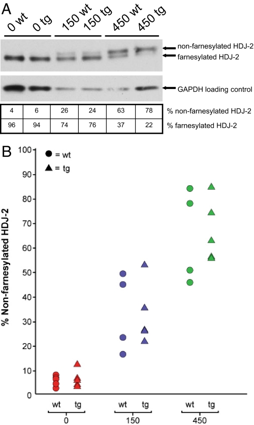
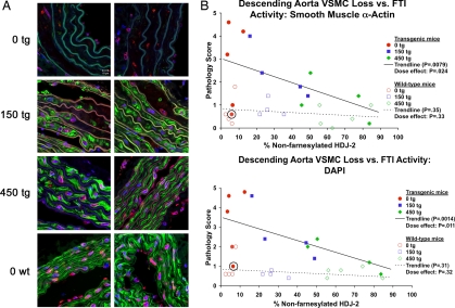
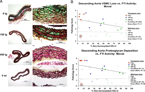
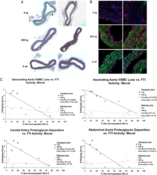
Hutchinson-Gilford progeria syndrome (HGPS) is the most dramatic form of human premature aging. Death occurs at a mean age of 13 years, usually from heart attack or stroke. Almost all cases of HGPS are caused by a de novo point mutation in the lamin A (LMNA) gene that results in production of a mutant lamin A protein termed progerin. This protein is permanently modified by a lipid farnesyl group, and acts as a dominant negative, disrupting nuclear structure. Treatment with farnesyltransferase inhibitors (FTIs) has been shown to prevent and even reverse this nuclear abnormality in cultured HGPS fibroblasts. We have previously created a mouse model of HGPS that shows progressive loss of vascular smooth muscle cells in the media of the large arteries, in a pattern that is strikingly similar to the cardiovascular disease seen in patients with HGPS. Here we show that the dose-dependent administration of the FTI tipifarnib (R115777, Zarnestra) to this HGPS mouse model can significantly prevent both the onset of the cardiovascular phenotype as well as the late progression of existing cardiovascular disease. These observations provide encouraging evidence for the current clinical trial of FTIs for this rare and devastating disease.


Conflict of interest statement

The authors declare no conflict of interest.

Figures

Fig. 1.
Fig. 1.
Oral FTI treatment leads to increased levels of the biomarker non-farnesylated HDJ-2. (A) Protein was extracted from mouse livers at the time of euthanasia. Western blotting of liver protein extracts demonstrates a dose-related increase in levels of non-farnesylated HDJ-2 (Upper) in comparison with farnesylated HDJ-2 (Lower) with FTI treatment. (B) All study mice are represented on a plot demonstrating the increasing levels of non-farnesylated HDJ-2 with increasing dose of FTI. Triangles represent transgenic G608G LMNA mice and circles represent non-transgenic WT controls.
Fig. 2.
Fig. 2.
FTI treatment begun at weaning prevents VSMC loss in G608G LMNA transgenic mice. (A) Analysis of the sections of the descending aortas of 9- to 12-month-old mice treated with 0, 150, or 450 mg/kg/day of FTI beginning at 1 month of age (blue, DNA; green, smooth muscle α-actin; red, lamins A/C; lamin A/C appears pink due to the overlay with the blue DAPI staining; white scale bar in upper left is 20 μm). The dose-dependent protective effect of the FTI is evident in the greater abundance of VSMCs displayed by the treated mice, as the 450 mg/kg/day treated (450 tg) mice appear virtually indistinguishable from the untreated WT mice (0 wt). (B) Scatter plots depicting average pathology scores (0–5 scale) given by five blinded observers scoring immunofluorescent (IF) descending aorta histological images from G608G LMNA transgenic mice for VSMC loss based upon smooth muscle α-actin staining loss and the loss of DAPI-positive nuclei versus levels of non-farnesylated HDJ-2. The trend lines demonstrate the significant prevention with increasing levels of FTI activity (P = 0.0079 and P = 0.0014). The circled mouse represents the one transgenic mouse in the studies that demonstrated epigenetic silencing of the transgene by quantitative RT-PCR and immunofluorescence and thus had no disease.
Fig. 3.
Fig. 3.
FTI treatment begun at weaning prevents VSMC loss and proteoglycan accumulation in G608G LMNA transgenic mice. (A) Analysis of the sections of the Movat-stained descending aortas of 9- to 12-month-old mice treated with 0, 150, or 450 mg/kg/day of FTI beginning at 1 month of age (red, VSMCs; green, proteoglycan; black, elastin layers). The dose-dependent protective effect is clearly seen in the greater cellularity and reduced proteoglycan staining in the treated transgenic animals (150 tg and 450 tg) in comparison with the untreated animals (0 tg). (B) Scatter plots depicting average pathology scores (0–5 scale) given by five blinded observers scoring the Movat descending aorta histological images from G608G LMNA transgenic mice for pathology in the forms of proteoglycan accumulation and VSMC loss versus levels of non-farnesylated HDJ-2. The circled mouse represents the one transgenic mouse in the studies that demonstrated epigenetic silencing of the transgene by quantitative RT-PCR and immunofluorescence and thus had no disease. The trend lines became significant upon removal of this outlier transgenic mouse from the analysis.
Fig. 4.
Fig. 4.
FTI treatment begun at 9 months of age prevents the late progression of VSMC loss and proteoglycan accumulation in G608G LMNA transgenic mice. (A) Movat-stained sections of the descending aortas of four 15-month-old transgenic mice treated with 0 or 450 mg/kg/day of FTI beginning at 9 months of age (red, VSMCs; green, proteoglycan; black, elastin layers). Whereas untreated 15-month-old transgenic mice display a complete lack of VSMCs and abundant proteoglycan accumulation as well as marked adventitial thickening (black arrowhead), treated mice appear virtually identical to age-matched untreated WT controls. (B) Immunofluorescence of the sections of the descending aortas of four 15-month-old mice treated with 0 or 450 mg/kg/day of FTI (blue, DNA; green, smooth muscle α-actin; red, lamins A/C; lamin A/C appears pink due to the overlay with the blue DAPI staining; white scale bar in upper left is 20 μm). Both sets of sections demonstrate complete protection in mice beginning treatment at the late age of 9 months. Untreated vessels are almost completely devoid of any VSMCs in the media, and proteoglycan has accumulated to replace them. In contrast, FTI-treated mice appear quite similar to untreated WT control mice with the vessel media well populated by VSMCs. (C) A sampling of scatter plots depicting average pathology scores (0–5 scale) given by five blinded observers scoring Movat descending aorta, ascending aorta, carotid artery, and abdominal aorta images from G608G LMNA transgenic mice treated from 9 to 15 months of age. The trend lines demonstrate the highly significant prevention trend with increasing levels of FTI activity in all plots.

References

    1. Meredith MA, et al. Phenotype and course of Hutchinson-Gilford progeria syndrome. N Engl J Med. 2008;358:592–604. - PMC - PubMed
    1. Eriksson M, et al. Recurrent de novo point mutations in lamin A cause Hutchinson-Gilford progeria syndrome. Nature. 2003;423:293–298. - PMC - PubMed
    1. De Sandre-Giovannoli A, et al. Lamin a truncation in Hutchinson-Gilford progeria. Science. 2003;300:2055. - PubMed
    1. Capell BC, Collins FS. Human laminopathies: nuclei gone genetically awry. Nat Rev Genet. 2006;7:940–952. - PubMed
    1. Dechat T, et al. . Alterations in mitosis and cell cycle progression caused by a mutant lamin A known to accelerate human aging. Proc Natl Acad Sci USA. 2007;104:4955–4960. - PMC - PubMed

MeSH terms

LinkOut - more resources