. 2008 Nov 21;3(11):711-22.
doi: 10.1021/cb800120t.

DcpS as a therapeutic target for spinal muscular atrophy

Affiliations

DcpS as a therapeutic target for spinal muscular atrophy

Jasbir Singh et al. ACS Chem Biol. .

Abstract

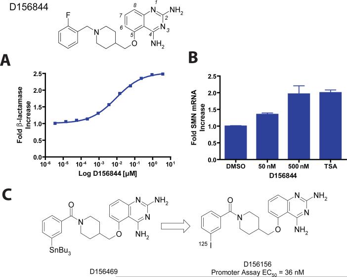
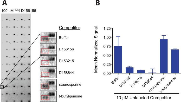
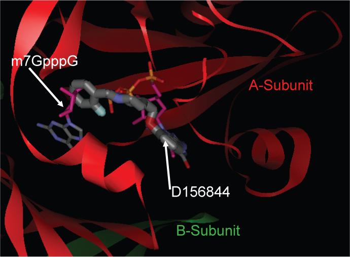
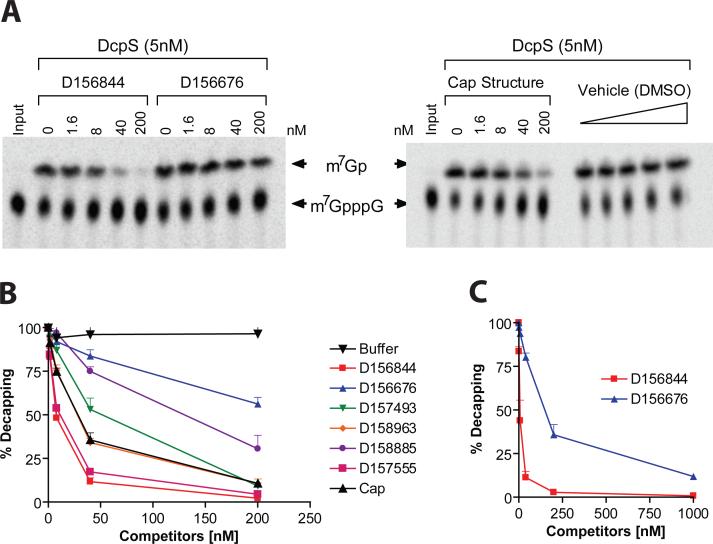
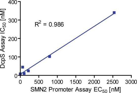
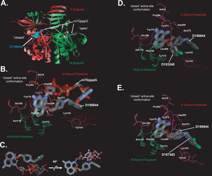
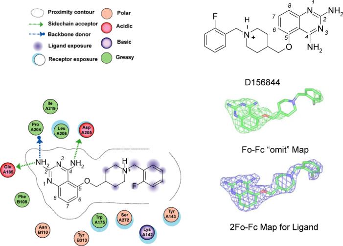
Spinal muscular atrophy (SMA) is caused by deletion or mutation of both copies of the SMN1 gene, which produces an essential protein known as SMN. The severity of SMA is modified by variable copy number of a second gene,SMN2, which produces an mRNA that is incorrectly spliced with deletion of the last exon. We described previously the discovery of potent C5-substituted quinazolines that increase SMN2 gene expression by 2-fold. Discovery of potent SMN2 promoter inducers relied on a cellular assay without knowledge of the molecular target. Using protein microarray scanning with a radiolabeled C5-substituted quinazoline probe, we identified the scavenger decapping enzyme, DcpS, as a potential binder. We show that the C5-substituted quinazolines potently inhibit DcpS decapping activity and that the potency of inhibition correlates with potency forSMN2 promoter induction. Binding of C5-substituted quinazolines to DcpS holds the enzyme in an open, catalytically incompetent conformation. DcpS is a nuclear shuttling protein that binds and hydrolyzes the m(7)GpppN mRNA cap structure and a modulator of RNA metabolism. Therefore DcpS represents a novel therapeutic target for modulating gene expression by a small molecule.


Figures

Figure 1
Figure 1. Structure of the C5-quinazoline D156844 and its effects on cellular SMN mRNA levels
The structure of D156844, a C5 substituted ether-based 2,4-diaminoquinazoline is shown with its 2-fluoro-benzyl piperidine substituent. Atoms of the quinazoline scaffold are numbered in italics. (A) Fold increase of β-lactamase in NSC34 cells treated with D156844 (mean ± SD, triplicate wells), EC50 = 9.1 nM (B) Fold increase of mouse SMN mRNA in NSC-34 cells treated with D156844, 100 nM trichostatin A (TSA) or DMSO (mean ± SD, n=9) (C) Synthesis of 125I labeled C5-quinazoline. D156469, a tin derivative was synthesized from D156156 as described in the Supplemental Text. An 125I sodium iodide /chloramine T procedure was used to displace the SnBu3 of D156469, yielding the desired radiolabeled C5-quinazoline.
Figure 2
Figure 2. Protein microarray identification of DcpS as a target for C5-substituted quinazolines
(A) Array images identifying DcpS as a potential D156156 target protein. Arrays comprised of 5,000 immobilized human proteins were probed with 125I-D156156 in the presence and absence of unlabeled competitors. Specific displacement of 125I-labeled D156156 (at 100 nM concentration) by bioactive, but distinct C5-quinazolines (unlabeled at 10 μM concentration) was observed. Duplicate features boxed in white correspond to the positional mapping reagent 125I-streptavidin binding to a biotinylated control protein. Duplicate features boxed in red correspond to DcpS. (B) Quantified competition binding data with 10 μM competitor normalized against signals arising from 125I-streptavidin binding to the biotinylated control protein. Standard deviations across the replicate assays are indicated.
Figure 3
Figure 3. Computational model for D156844 binding to the closed active site conformation of DcpS
An energy minimized conformer of D156844 was computationally docked into the closed active site conformation of human DcpS (PDB ID:1ST0). The final pose for D156844 (thick stick) is shown superimposed on the structure of m7GpppG (thin stick), both bound into the asymmetric DcpS homodimer (red: A-subunit; green: B-subunit).
Figure 4
Figure 4. Inhibition of DcpS decapping activity by C5-substituted quinazolines
(A) Radiolabeled m7Gp*pppG was incubated with purified human DcpS (5nM) in the presence of increasing concentrations of C5-quinazolines (D156844 or D156676), a positive control for DcpS inhibition (Cap structure), or a negative vehicle (DMSO) control. The reaction products were separated by thin layer chromatography and visualized with a Phosphorimager. (B) Data for the relative decapping rate vs. concentration of compounds (nM). Structures of the compounds are shown in Table 1. (C) Inhibition of endogenous DcpS decapping activity in 20μg of human K562 erythroleukemia cellular extract with the D156844 or D156676 C5-quinazolines.
Figure 5
Figure 5
Linear correlation between SMN2 promoter inducing activity and DcpS decapping inhibition.
Figure 6
Figure 6. Crystal Structures of DcpS Bound to C5-quinazolines
(A) Superposition of DcpS with bound D156844 and m7GpppG. The structure of DcpS bound to D156844 (cyan CPK space filling model) to human DcpS (pink Subunit A, and light green Subunit B) has been superimposed onto the previously reported structure of human DcpS (dark red Subunit A, and dark green Subunit B) with m7GpppG (stick model) bound to both the closed (left side) and open (right side) active site regions (PDB ID:1ST0) . The superposition illustrates that D156844 is bound only in closed active site conformation state, while m7GpppG is capable of binding into both the open and closed active site pockets. The DcpS structures with D156844 and m7GpppG bound superimpose with and RMSD on all atoms of 1.1 Å indicating that the conformations of the two are very similar. (B) Superposition of D156844 and m7GpppG in the DcpS active site. The structure of DcpS bound to D156844 (labeled stick model) to human DcpS (pink Subunit A, and light green Subunit B) has been superimposed onto the previously reported structure of human DcpS (dark red Subunit A, and dark green Subunit B) with m7GpppG (labeled stick model) bound active site regions (PDB ID:1ST0) . The “closed” active-site conformation of DcpS is shown. Amino acid side chains are numbered and the ligands are identified. The 2−4 diamino quinazoline of for D156844 ligand adopts an identical binding mode as the m7G group of m7GpppG in DcpS. The C5-substituent of D156844 takes on a different conformation in binding to DcpS as compared to the pppG portion of the m7GpppG. Side chains which take on significantly different conformations between the two structures are Tyr273 and Lys142. (C) Comparison of the binding pose of D156844 to m7GpppG cap substrate. The DcpS protein structures of our C5-quinazoline bound structure and that of the m7GpppG bound structure (PDB ID:1ST0) were superimposed with COOT . The superposition the DcpS protein structures revealed that the quinazoline moiety D156844 (thick ball and stick) overlays nicely with that of the m7G moiety of m7GpppG (thin ball and stick) of the 1ST0 structure. In contrast, the C5 substituent of the quinazoline follows a different trajectory in the binding pocket compared to the pppG appendage of m7GpppG. The two images are rotated 90 degrees with respect to each other and the protein has been removed. (D) Superposition of DcpS with bound D156844 and D153249. The “closed” active-site conformation of DcpS is shown in superposition mode for the DcpS co-crystal structures with D156844 (light pink for A subunit residues and light green for B subunit residues) and D153249 (dark pink for A subunit residues and dark green for B subunit residues). Amino acid side chains are numbered and the ligands are identified. The 2−4 diamino quinazoline of for each ligand adopts an identical binding mode in DcpS. The C5-substituent of each ligand takes on a different conformation in binding to DcpS. (E) Superposition of DcpS with bound D156844 and D157493. The “closed” active-site conformation of DcpS is shown in superposition mode for the DcpS co-crystal structures with D156844 (light pink for A subunit residues and light green for B subunit residues) and D157493 (dark pink for A subunit residues and dark green for B subunit residues). Amino acid side chains are numbered and the ligands are identified. The 2−4 diamino quinazoline of for each ligand adopts an identical binding mode in DcpS. The C5-substituent of each ligand takes on a different conformation in binding to DcpS.
Figure 7
Figure 7. Ligand Interaction Figure of D156844 bound to Human DcpS “Closed” Active Site
Left Panel. DcpS amino acid interactions with D156844 are depicted schematically for the closed active site. The “A” and “B” subunit residues are listed with “A” or “B” prefixing the residue number. The figure was generated by the MOE software package (Chemical Computing Group, Montreal, Canada), with legend shown. Upper Right Panel. The chemical structure of D156844. Middle Right Panel. Green wire, 3.0 sigma positive electron density(|Fo|-|Fc|) of omit map of D156844 bound in the closed active site of human DcpS. Lower Right Panel. Blue wire, final 2|Fo|-|Fc| electron density map of D156844 bound in the closed active site of human DcpS.

References

    1. Shiau AK, et al. The structural basis of estrogen receptor/coactivator recognition and the antagonism of this interaction by tamoxifen. Cell. 1998;95:927–37. - PubMed
    1. Kliewer SA, Lehmann JM, Milburn MV, Willson TM. The PPARs and PXRs: nuclear xenobiotic receptors that define novel hormone signaling pathways. Recent Prog Horm Res. 1999;54:345–67. discussion 367−8. - PubMed
    1. Pearn J. Incidence, prevalence, and gene frequency studies of chronic childhood spinal muscular atrophy. J Med Genet. 1978;15:409–13. - PMC - PubMed
    1. Roberts DF, Chavez J, Court SD. The genetic component in child mortality. Arch Dis Child. 1970;45:33–8. - PMC - PubMed
    1. Lefebvre S, et al. Identification and characterization of a spinal muscular atrophy-determining gene. Cell. 1995;80:155–65. - PubMed

Publication types

MeSH terms