. 2008 Oct 7:8:288.
doi: 10.1186/1471-2407-8-288.

High-resolution array CGH clarifies events occurring on 8p in carcinogenesis

Affiliations

High-resolution array CGH clarifies events occurring on 8p in carcinogenesis

Susanna L Cooke et al. BMC Cancer. .

Abstract

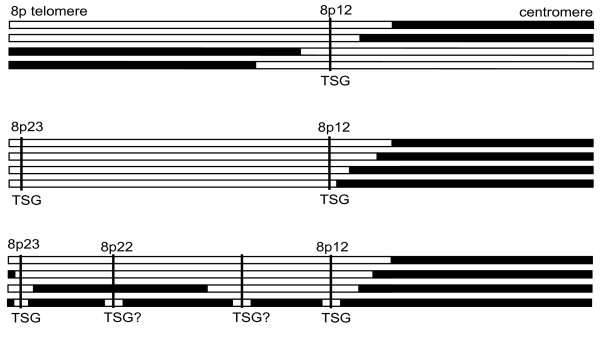
Background: Rearrangement of the short arm of chromosome 8 (8p) is very common in epithelial cancers such as breast cancer. Usually there is an unbalanced translocation breakpoint in 8p12 (29.7 Mb - 38.5 Mb) with loss of distal 8p, sometimes with proximal amplification of 8p11-12. Rearrangements in 8p11-12 have been investigated using high-resolution array CGH, but the first 30 Mb of 8p are less well characterised, although this region contains several proposed tumour suppressor genes.

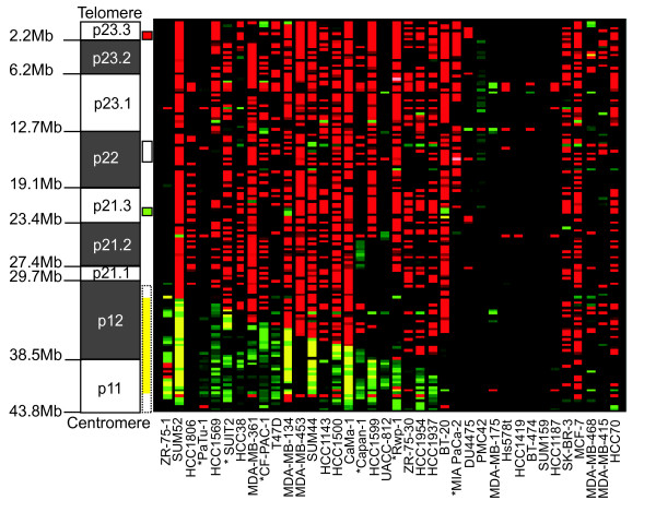
Methods: We analysed the whole of 8p by array CGH at tiling-path BAC resolution in 32 breast and six pancreatic cancer cell lines. Regions of recurrent rearrangement distal to 8p12 were further characterised, using regional fosmid arrays. FISH, and quantitative RT-PCR on over 60 breast tumours validated the existence of similar events in primary material.

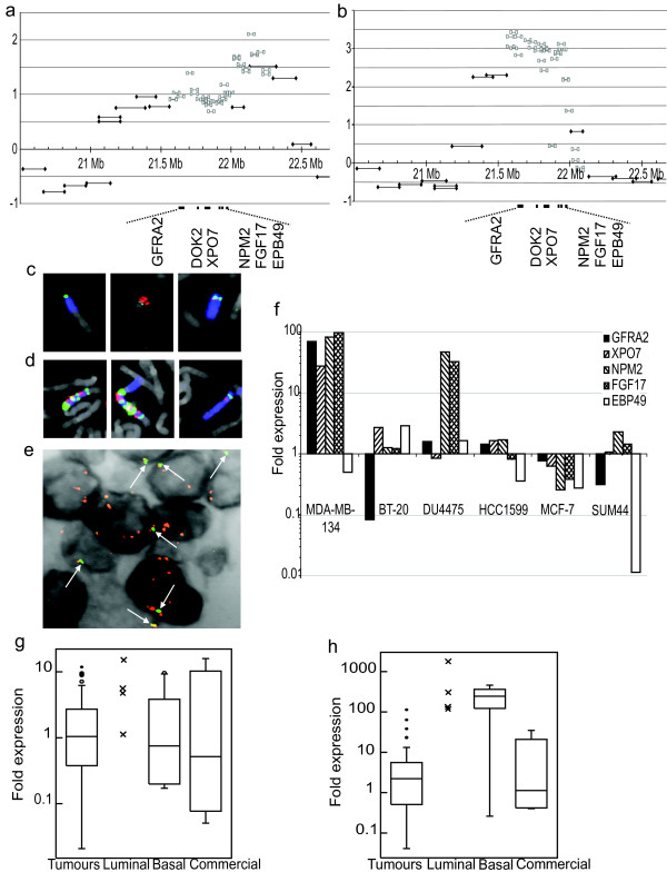
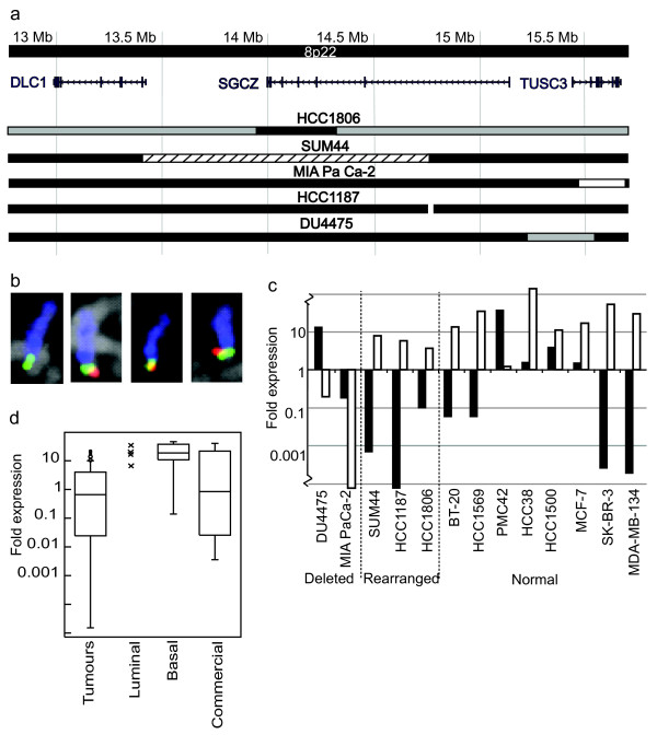
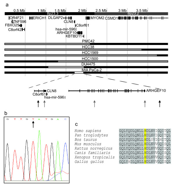
Results: We confirmed that 8p is usually lost up to at least 30 Mb, but a few lines showed focal loss or copy number steps within this region. Three regions showed rearrangements common to at least two cases: two regions of recurrent loss and one region of amplification. Loss within 8p23.3 (0 Mb - 2.2 Mb) was found in six cell lines. Of the genes always affected, ARHGEF10 showed a point mutation of the remaining normal copies in the DU4475 cell line. Deletions within 12.7 Mb - 19.1 Mb in 8p22, in two cases, affected TUSC3. A novel amplicon was found within 8p21.3 (19.1 Mb - 23.4 Mb) in two lines and one of 98 tumours.

Conclusion: The pattern of rearrangements seen on 8p may be a consequence of the high density of potential targets on this chromosome arm, and ARHGEF10 may be a new candidate tumour suppressor gene.


Figures

Figure 1
Figure 1
Array CGH of 8p in 38 cancer cell lines. The ideogram shows the short arm of chromosome 8 with the position of the chromosome bands indicated on the left and regions of recurrent change represented by coloured boxes on the right of the ideogram. Previously identified positions of recurrent rearrangement (8p11-12) are indicated by a dashed outline. The novel regions identified by this study are indicated by a solid outline. For these summary boxes red indicates a common region of loss in 8p23.3, green a common region of gain/amplification in 8p21.3 and white indicates a region with various rearrangements including gain, loss and breakpoints in 8p22. Array CGH results are displayed as a heat map using CGHAnalyzer (, [64]). The first twenty-four lines show a typical pattern of 8p12 rearrangement with breakpoints, distal loss and occasionally proximal amplification. The next four lines have atypical patterns of 8p rearrangement. The final ten lines show no copy number changes within 8p but the last five have lost the whole of 8p with respect to 8q. Red, loss relative to the ploidy of the cell line; Black, no change; Green, gain; Yellow, high level amplification; Grey, no copy number data; * indicates a pancreatic line.
Figure 2
Figure 2
Analysis of the 8p21.3 amplicon. (a) and (b) BAC and fosmid array CGH of the 8p21.3 amplicons in BT-20 and MDA-MB-134 respectively. (a) BT-20 showed amplification between BAC clones RP11-419L22 (21.13 Mb; positions are given as midpoints on NCBI Build 36) and RP11-582J16 (22.50 Mb). (b) MDA-MB-134 showed amplification between BAC clones RP11-458H21 (21.28 Mb) and RP13-600L4 (22.04 Mb). The distal edges of both amplicons fell in a region containing no known genes but extended proximally into a gene-dense region. Genes within the overlap are marked. Log2 ratio of the fluorescence intensity is plotted against position on 8p. Grey squares, fosmids; Black diamonds, BACs. (c) FISH of the three chromosomes containing material from the 8p21.3 amplicon in BT-20. Blue, chromosome 8 paint; Green, RP11-459H21 (21.29 Mb); Red, RP11-235B11 (22.38 Mb). (d) FISH of the three chromosomes containing material from the 8p21.3 and 8p12 amplicons in MDA-MB-134. Blue, chromosome 8 paint; Green, BAC RP11-135I5 (21.49 Mb); Red, RP11-104D16 (40.25 Mb). (e) FISH of the 8p21.3 amplicon on a primary breast tumour paraffin section. Red, BAC pool positioned in the amplicon (centred at 21.9 Mb); Green (also indicated by arrows), BAC pool distal to the amplicon (centred at 19.5 Mb); Grey, inverted DAPI. (f) Expression levels of the five genes included in both the BT-20 and MDA-MB-134 amplicons, shown on the y-axis as a log10 scale of -fold expression compared to normal breast cell line HB4a and normalised to GAPDH as an internal control. Expression levels of (g) FGF17 and (h) NPM2 in primary tumours and normal controls. Crosses, purified luminal samples; Open circles, possible outliers (value more than 1.5× inter-quartile range above the third quartile); Filled circles, outliers (value more than 3× inter-quartile range above the third quartile).
Figure 3
Figure 3
Analysis of 8p22 rearrangements. (a) Rearrangements between 12.9 Mb and 15.6 Mb in 8p22. Black bar, normal copy number for 8p; Grey, heterozygous loss; White with black outline, homozygous loss; White with no outline, breakpoint of reciprocal translocation; Black hatching, gain. (b) FISH showing the deletion of the TUSC3 promoter on one of the four copies of chromosome 8 in DU4475. Blue, chromosome 8 paint; Green, RP11-480M18 (15.0 Mb); Red, RP11-314P10 (15.6 Mb). (c) DLC1 (black bars) and TUSC3 (white bars) expression levels relative to normal breast line HB4a. Note TUSC3 expression is decreased in MIA PaCa-2 and DU4475, the two cell lines with deletions of this gene. (d) TUSC3 expression in primary breast tumours and various normal breast samples (see Methods). TUSC3 expression is lower in a subset of tumours than in any control group.
Figure 4
Figure 4
Analysis of losses in 8p23.3. (a) Losses between 3.7 Mb and the telomere. Black bar, normal copy number for 8p; Grey, heterozygous loss; White with black outline, homozygous loss. Black arrows, position of primer pairs that amplify a product from MIA PaCa-2 genomic DNA i.e. lie outside the homozygous deletion; Grey arrows, position of primer pairs that do not amplify a product from MIA PaCa-2 genomic DNA. (b) Sequencing trace showing the point mutation in exon 19 of ARHGEF10. The normal sequence has a cytosine at the position indicated by the arrow. (c) Conservation of ARHGEF10. Yellow, the residue that is converted from a histidine to tyrosine by the point mutation in DU4475; Grey, conserved residues.
Figure 5
Figure 5
A parsimonious model of 8p rearrangements. The different patterns of 8p rearrangement that would be expected in a sample of tumours based on the number and position of tumour suppressor genes (not to scale). Top is the pattern of rearrangements expected if there is a single tumour suppressor gene located in 8p12. Middle is the pattern of rearrangements predicted if there were at least two tumour suppressor genes located on 8p, one in 8p12 and one located close to the telomere in 8p23.3. This fits the common overall pattern of breaks in 8p12 with loss of distal 8p. Bottom is a representation of some of the atypical 8p rearrangements found in cancer cell lines including DU4475, HCC1500 and PMC42. These can be explained by the presence of other tumour suppressor genes in between those in 8p12 and 8p23.3; our results suggest at least one, at 8p22. Since each of these tumour suppressor genes is only lost in a proportion of cases a complex pattern of rearrangements encompassing some or all of these regions results. Loss of the whole of distal 8p will be the most common rearrangement as it removes all tumour suppressor genes as the result of a single event.

References

    1. Tirkkonen M, Tanner M, Karhu R, Kallioniemi A, Isola J, Kallioniemi OP. Molecular cytogenetics of primary breast cancer by CGH. Genes Chromosomes Cancer. 1998;21:177–184. doi: 10.1002/(SICI)1098-2264(199803)21:3<177::AID-GCC1>3.0.CO;2-X. - DOI - PubMed
    1. Loo LW, Grove DI, Williams EM, Neal CL, Cousens LA, Schubert EL, Holcomb IN, Massa HF, Glogovac J, Li CI, Malone KE, Daling JR, Delrow JJ, Trask BJ, Hsu L, Porter PL. Array comparative genomic hybridization analysis of genomic alterations in breast cancer subtypes. Cancer Res. 2004;64:8541–8549. doi: 10.1158/0008-5472.CAN-04-1992. - DOI - PubMed
    1. Davidson JM, Gorringe KL, Chin SF, Orsetti B, Besret C, Courtay-Cahen C, Roberts I, Theillet C, Caldas C, Edwards PA. Molecular cytogenetic analysis of breast cancer cell lines. Br J Cancer. 2000;83:1309–1317. doi: 10.1054/bjoc.2000.1458. - DOI - PMC - PubMed
    1. Bashyam MD, Bair R, Kim YH, Wang P, Hernandez-Boussard T, Karikari CA, Tibshirani R, Maitra A, Pollack JR. Array-based comparative genomic hybridization identifies localized DNA amplifications and homozygous deletions in pancreatic cancer. Neoplasia. 2005;7:556–562. doi: 10.1593/neo.04586. - DOI - PMC - PubMed
    1. Emi M, Fujiwara Y, Nakajima T, Tsuchiya E, Tsuda H, Hirohashi S, Maeda Y, Tsuruta K, Miyaki M, Nakamura Y. Frequent loss of heterozygosity for loci on chromosome 8p in hepatocellular carcinoma, colorectal cancer, and lung cancer. Cancer Res. 1992;52:5368–5372. - PubMed

Publication types