. 2008 Oct 7:9:58.
doi: 10.1186/1471-2172-9-58.

Transglutaminase activity in the hematopoietic tissue of a crustacean, Pacifastacus leniusculus, importance in hemocyte homeostasis

Affiliations

Transglutaminase activity in the hematopoietic tissue of a crustacean, Pacifastacus leniusculus, importance in hemocyte homeostasis

Xionghui Lin et al. BMC Immunol. .

Abstract

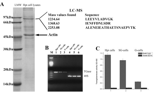
Background: Transglutaminases (TGases) form a group of enzymes that have many different substrates and among the most well known are fibrin for Factor XIIIa and the clotting protein in crustaceans. We also found that TGase is an abundant protein in the hematopoietic tissue (Hpt) cells of crayfish and hence we have studied the possible function of this enzyme in hematopoiesis.

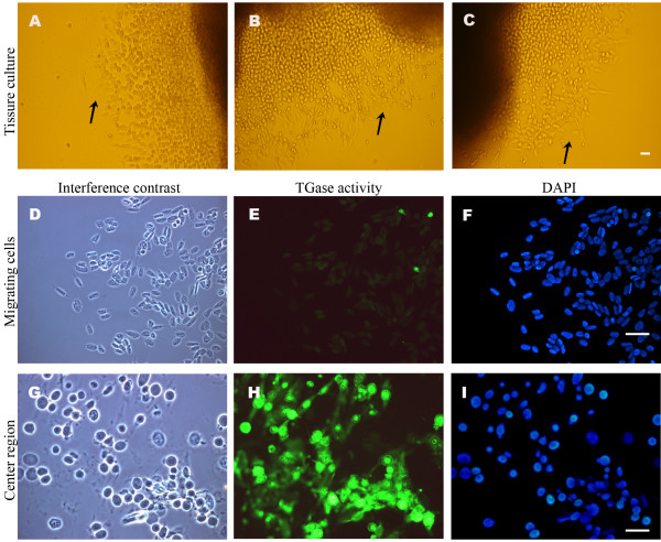
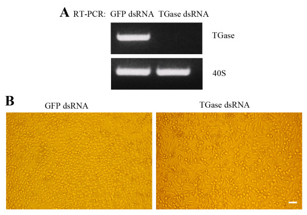
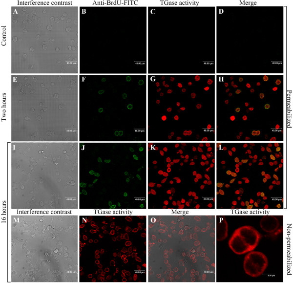
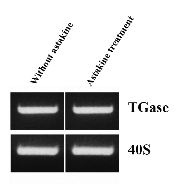
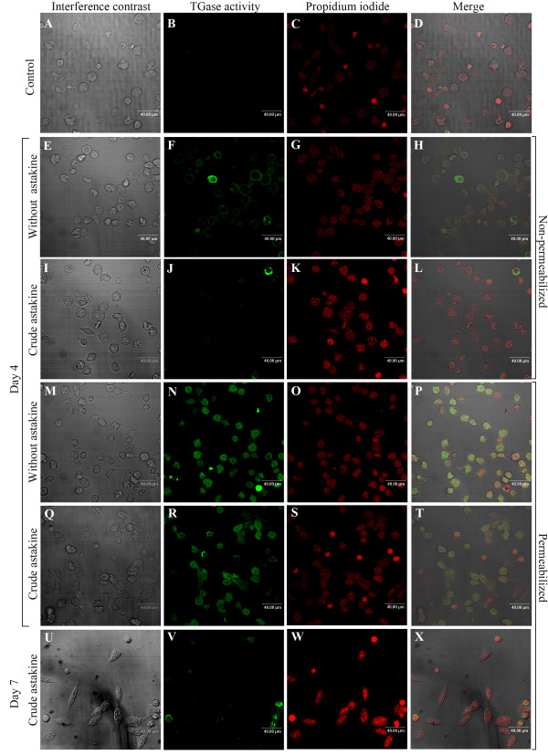
Results: TGase is one of the most abundant proteins in the Hpt and its mRNA expression as well as enzyme activity is very high in the Hpt cells, lesser in the semi-granular hemocytes and very low in the granular cells. In cultured hematopoietic tissues, high activity was present in cells in the centre of the tissue, whereas cells migrating out of the tissue had very low TGase activity. RNAi experiments using dsRNA for TGase completely knocked down the transcript and as a result the cell morphology was changed and the cells started to spread intensely. If astakine, a cytokine directly involved in hematopoiesis, was added the cells started to spread and adopt a morphology similar to that observed after RNAi of TGase. Astakine had no effect on TGase expression, but after a prolonged incubation for one week with this invertebrate cytokine, TGase activity inside and outside the cells was completely lost. Thus it seems as if astakine addition to the Hpt cells and RNAi of TGase in the cell culture will lead to the same results, i.e. loss of TGase activity in the cells and they start to differentiate and spread.

Conclusion: The results of this study suggest that TGase is important for keeping the Hpt cells in an undifferentiated stage inside the hematopoietic tissue and if expression of TGase mRNA is blocked the cells start to differentiate and spread. This shows a new function for transglutaminase in preventing hematopoietic stem cells from starting to differentiate and migrate into the hemolymph, whereas their proliferation is unaffected. Astakine is also important for the hematopoiesis, since it induces hemocyte synthesis in the Hpt but now we also show that it in some unknown way participates in the differentiation of the Hpt cells.


Figures

Figure 1
Figure 1
TGase is an abundant protein in Hpt cells. A. Hpt cell lysate separated by 12.5% SDS-PAGE. The main protein band around 90 kDa was confirmed to be the crayfish TGase by LC-MS. The strong band at 50 kDa (arrow) was confirmed to be actin. B. TGase transcript levels in Hpt cells and in separated circulating SG and G hemocytes analyzed by RT-PCR. Control transcript is 40S ribosomal protein. Lane 1, Hpt cell TGase; lane 2, SG-cell TGase; lane 3, G-cell TGase; lane 4, Hpt cells 40S; lane 5, SG-cell 40S; lane 6, G-cell 40S; M is 100 bp DNA marker. C. TGase activity in Hpt cell lysate and in lysate from separated SG or G hemocytes. The amount of incorporated 5' (biotinamido) pentylamine was quantified by measuring the absorbance at 450 nm in a plate reader. Mean ± SD of 3 separate experiments.
Figure 2
Figure 2
The promoter region of TGase contains GATA motifs. Schematic diagram of the 5'-flanking regulatory sequences and distribution of GATA motif in crayfish TGase and some human TGase genes.
Figure 3
Figure 3
TGase activity is low in cells migrating out of the Hpt. Hematopoietic tissues were cultured on coverslips for one week, and labelled with 1 mM BPNH2 overnight. A-C. Migrating (spread) cells (arrows) can be seen at distance from the centre of the tissue; D-F. TGase activity in migrating cells; G-I. TGase activity in cells close to the centre of the tissue. Interference contrast (D, G); TGase activity (E, H); Nuclear staining was performed with DAPI (F, I). Bar is 40 μm.
Figure 4
Figure 4
RNA interference of TGase in cultured Hpt cells. A. TGase expression analyzed by RT-PCR, five days after transfection with TGase dsRNA or GFP dsRNA (as control). 40 S ribosomal protein was used as a control transcript. B. Morphology change of Hpt cells transfected with dsRNA for TGase. Left, control cells transfected with GFP dsRNA; right, cells transfected with TGase dsRNA. Bar is 40 μm.
Figure 5
Figure 5
TGase activity of HSCs does not prevent its proliferation. Hpt cells were cultured on coverslips, and maintained in six well plates. The cells were labeled with 1 mM 5-(biotinamindo)-pentylamine (BPNH2; Molecular Probes, Pierce) as a substrate for TGase and 10 μM BrdU (Sigma) for 2 or 16 hours. The in situ TGase activity was stained by streptavidin-Cy5, BrdU incorporation assay was measured by staining with anti-BrdU-FITC. A-D. Control cells incubated without BrdU and BPNH2.E-H. Cells incubated with BrdU and BPNH2 for 2 hours. I-L. Cells incubated with BrdU and BPNH2 for 16 hours. M-P. Cell surface TGase activity detected in non-permeabilized cells after incubation with BPNH2 for 16 hours.
Figure 6
Figure 6
Astakine does not affect TGase mRNA expression. TGase expression analyzed by RT-PCR, four days after culture in the presence or absence of astakine. 40 S ribosomal protein was used as a control transcript.
Figure 7
Figure 7
Astakine treatment results in decreased TGase activity in the Hpt cells. Hpt cells were seeded on coverslips, and maintained in culture dishes, and the medium supplied was added with or without astakine (a crayfish prokineticin domain-containing protein) [21]. At day 4 (E-T) and day 7 (U-X), the cells were incubated with 1 mM BPNH2 in medium two hours at room temperature, and visualized using the streptavidin-FITC conjugate. Nuclear staining was performed with propidium iodide. A-D. Non-permeabilized Hpt cells cultured for 4 days without BPNH2 were used as a staining control. E-H. Non-permeabilized Hpt cells cultured for 4 days without astakine showing surface TGase activity. I-L. Non-permeabilized Hpt cells cultured for 4 days with astakine showing no surface TGase activity. M-P. Permeabilized Hpt cells cultured for 4 days without astakine. Q-T. Permeabilized Hpt cells cultured for 4 days with astakine. U-X. Permeabilized Hpt cells cultured for 7 days with astakine showing no TGase activity. Bar is 40 μm.

References

    1. Ichinose A, Bottenus RE, Davie EW. Structure of transglutaminases. J Biol Chem. 1990;265:13411–13414. - PubMed
    1. Steinert PM, Kartasova T, Marekov LN. Biochemical Evidence That Small Proline-rich Proteins and Trichohyalin Function in Epithelia by Modulation of the Biomechanical Properties of Their Cornified Cell Envelopes. J Biol Chem. 1998;273:11758–11769. doi: 10.1074/jbc.273.19.11758. - DOI - PubMed
    1. Ho KC, Quarmby VE, French FS, Wilson EM. Molecular cloning of rat prostate transglutaminase complementary DNA. The major androgen-regulated protein DP1 of rat dorsal prostate and coagulating gland. J Biol Chem. 1992;267:12660–12667. - PubMed
    1. Wang R, Liang Z, Hall M, Söderhäll K. A transglutaminase involved in the coagulation system of the freshwater crayfish, Pacifastacus leniusculus. Tissue localisation and cDNA cloning. Fish Shellfish Immunol. 2001;11:623–637. doi: 10.1006/fsim.2001.0341. - DOI - PubMed
    1. Osaki T, Okino N, Tokunaga F, Iwanaga S, Kawabata S-i. Proline-rich Cell Surface Antigens of Horseshoe Crab Hemocytes Are Substrates for Protein Cross-linking with a Clotting Protein Coagulin. J Biol Chem. 2002;277:40084–40090. doi: 10.1074/jbc.M206773200. - DOI - PubMed

Publication types

Substances

LinkOut - more resources