Review
. 2009 Jan 15;384(2):197-206.
doi: 10.1016/j.ab.2008.09.020. Epub 2008 Sep 18.

Tubulin proteomics: towards breaking the code

Affiliations
Review

Tubulin proteomics: towards breaking the code

Pascal Verdier-Pinard et al. Anal Biochem. .

Abstract

Since the discovery of tubulin as the major component of microtubules over 40 years ago, its diversity of forms has raised a continuum of fundamental questions about its regulation and functions in a variety of organisms across phyla. Its high abundance in the brain or in specialized organelles such as cilia has allowed early characterization of this important target for anticancer drugs. However, it was only when matrix-assisted laser desorption ionization and electrospray ionization mass spectrometry technologies became available in the late 1980's that the full complexity of tubulin expression patterns became more obvious. This contributed in a major way to the idea that due to increasing and conserved tubulin heterogeneity during evolution, a tubulin code read by microtubule associated proteins might exist and be of functional significance. We review here the merging of recent genetic and cell biology studies with proteomics to decipher this code and illustrate some of the tubulin proteomic approaches with new data generated in our laboratories.


Figures

Figure 1
Figure 1. Mass spectrometry analysis of CNBr C-terminal peptides from α- or β-tubulins-separated by SDS-PAGE
Tubulin was isolated from A549 or HMEC-1 cells and separated on a 10 % acrylamide gel containing 2 M urea at pH 9.5. After Coomassie staining, the gel band containing either α-tubulin (left panels) or β-tubulin (right panels) was incubated with CNBr and formic acid (FA) and analyzed by MALDI-TOF MS in the negative mode. Top and middle panels, two independent preparations of CNBr peptides from tubulin isolated from A549 cells; bottom panels, CNBr peptides from tubulin isolated from HMEC-1 cells; mono- and bi- formylated peptides are labeled by one star (+28 Da) and two stars (+56 Da), respectively; monoglutamylated peptides are labeled with filled symbols (+129 Da); peptides from the different tubulin isotypes are indicated as follow: α1C (□), detyrosinated α1B (formula image
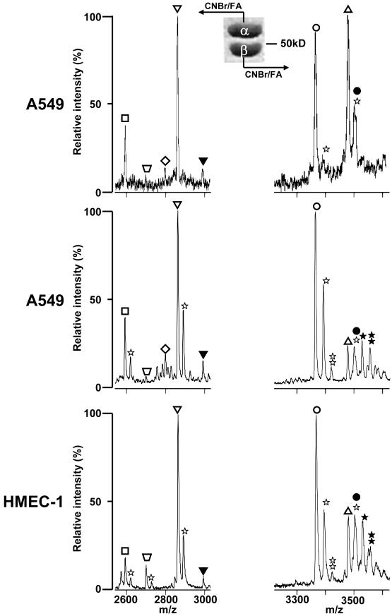
, −163 Da), tyrosinated α4A (◇, +163 Da), α1B (∇), βI (◯), βIVb (△). The assignment of each m/z peak for β-tubulin is an interpretation based on the CNBr C-terminal peptides of βI- and βIVb-tubulin and their monoformylated derivatives.
Figure 2
Figure 2. Mass spectrometry analysis of CNBr C-terminal peptides from IEF-separated tubulin isotypes from A549 and A549.EpoB40 cells
A, Schematic representation of a portion of an IPG strip (pH 4.5–5.5) with predicted positions of tubulin isotypes depicted as vertical black bands starting with monoglutamylated βI-tubulin (band 1, pI: 4.76) and finishing with α1C-tubulin (band 8, pI: 4.96). Bands 2 to 7 represent βI-, βIVb-, monoglutamylated βIII-, βIII, monoglutamylated α1B-, tyrosinated α4A/α1B- and α1C-tubulin, respectively. On Coomassie-stained IEF gels, the black indicate the position of wild type and mutated βI-tubulin from A549 and A549.EpoB40, respectively. The white arrow and the grey arrow indicate the focalization position of βII-and monoglutamylated mutated βI-tubulin, respectively. B, the bands corresponding to mutated βI-tubulin (left panel) and βII tubulin (right panel)were cut out and the protein was cleaved by CNBr. Peptides were analyzed by MALDI-TOF MS in the negative mode and only the band expected to contain βII-tubulin produced a small m/z peak (3467.71) corresponding to the C-terminus of βII-tubulin. Another m/z peak at 3496.50 corresponded to contaminating monoglutamylated βI tubulin (βIglu1). Maximal intensity is 114 for left spectrum vs 30 for right spectrum.
Figure 3
Figure 3. High resolution 2D-electrophoresis tubulin isotypes in Taxol-stabilized microtubules from HMEC-1, endothelial cells
Tubulin was isolated from HMEC-1 cells as described in the text. Tubulin isotypes were separated in the first dimension on a 24 cm IPG-strip pH 4.5–5.5 (pH 4.5–5.0 portion shown). The second dimension was run on a 10% acrylamide gel and proteins were blotted on a nitrocellulose membrane. The membrane was probed with antibodies against the indicated isotypes and stripped between each antibody. Alignment of blots was performed using the actin spots (not displayed). An anti-human βV-tubulin rabbit polyclonal antibody was produced and specifically labeled a spot on the acidic side of βI-tubulin as predicted. Insets show equivalent blots for α-, βI- and βII-tubulins in A549 cells; the most acidic spot for βI-tubulin corresponds to its monoglutamylated form.
Figure 4
Figure 4. Relative quantitation of total tubulin expression by SILAC
A, relative intensities of the m/z peaks of β-tubulin peptide β242-251 were obtained from SILAC experiments using d0- or d3-Leu in cell culture medium. The two leucine residues present in the β242-251 peptide are underlined and a difference of 6 Da is observed between the d0-β242-251 and d3-β242-251 m/z peaks. B, relative quantitation of total β-tubulin expression obtained after deisotoping of m/z peaks and averaging of relative intensities of m/z peaks in spectra from different β-tubulin peptides containing Leu. Tubulin was isolated and analyzed by MALDI-TOF MS from the following experiments as indicated on the top of mass spectra in A and under each bar of the graph in B: MDA-MD-231.K20T cells were cultured in the presence of d0-Leu after removal of normal culture medium containing 20 nM Taxol and one passage in Taxol-free medium. MDA-MD-231 cells were cultured in the presence of d3-Leu; MDA-MD-231 cells were maintained in the presence of d0-Leu and treated with 0.5 nM Taxol for 24 h, and untreated MDA-MD-231 cells were maintained in the presence of d3-Leu; MDA-MD-231 cells were maintained in the presence of d0-Leu and treated with 1.0 nM Taxol for 24 h, and untreated MDA-MD-231 cells were maintained in the presence of d3-Leu.

References

    1. Honoré S, Pasquier E, Braguer D. Understanding microtubule dynamics for improved cancer therapy. Cell Mol Life Sci. 2005;62:3039–56. - PMC - PubMed
    1. Orr GA, Verdier-Pinard P, McDaid H, Horwitz SB. Mechanisms of Taxol resistance related to microtubules. Oncogene. 2003;22:7280–95. - PMC - PubMed
    1. Rodriguez OC, Schaefer AW, Mandato CA, Forscher P, Bement WM, Waterman-Storer CM. Conserved microtubule-actin interactions in cell movement and morphogenesis. Nat Cell Biol. 2003;5:599–609. - PubMed
    1. Bonnet C, Boucher D, Lazereg S, Pedrotti B, Islam K, Denoulet P, Larcher JC. Differential binding regulation of microtubule-associated proteins MAP1A, MAP1B, and MAP2 by tubulin polyglutamylation. J Biol Chem. 2001;276:12839–48. - PubMed
    1. Boucher D, Larcher JC, Gros F, Denoulet P. Polyglutamylation of tubulin as a progressive regulator of in vitro interactions between the microtubule-associated protein Tau and tubulin. Biochemistry. 1994;33:12471–7. - PubMed

Publication types

LinkOut - more resources