

doi: 10.1021/pr800551m.
Epub 2008 Oct 8.
Proteomic analysis of the hyaloid vascular system regression during ocular development
Affiliations
- PMID: 18841878
- PMCID: PMC2662928
- DOI: 10.1021/pr800551m
Item in Clipboard
Proteomic analysis of the hyaloid vascular system regression during ocular development
J Proteome Res.
2008 Nov.
Abstract
We describe a proteomic approach to investigate the differential protein expression patterns and identify the physiologically relevant angiogenic and antiangiogenic factors involved in the hyaloid vascular system regression. Differentially expressed proteins were identified using two-dimensional gel electrophoresis followed by nanoflow chromatography coupled with tandem mass spectrometry. These proteins are expected to provide insight as to their function in the early maintenance and eventual regression of the hyaloid vascular system.
Conflict of interest statement
Commercial relationship and financial interest: None
Figures
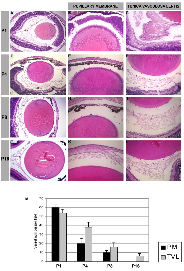
Hematoxylin-eosin staining of mouse eyes at different stages of development: P1 (A), P4 (D), P8 (G), and P16 (J), magnification (10x). Enlargement of the anterior part of the mouse lens with pupillary membrane (PM) vessels, magnification (20x) (B, E, H, K). Posterior part of the lens with the tunica vasculosa lentis (TVL) and the vasa hyaloidea propria (VHP), magnification (20x) (C, F, I, L). (M) Graph showing the vessel number per field at magnification (20x). (N) Graph showing the mean spot volume in pixels. (O) Graph showing the fold increase/decrease of each time point over P1.
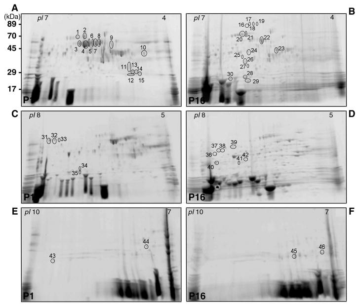
Representative 2-DE gels of proteins obtained from the lens and vitreous of P1 mouse (A, C, E) and P16 mouse (B, D, F) using IPG strips with pH range 4–7 (A, B), 5–8 (C, D), 7–10 (E, F). The proteins excised for analysis and identification by MS are marked with numbers from 1 to 46.
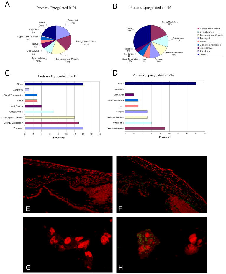
A–D: Pie-chart/frequency bar graphs showing the summary of protein classes that were upregulated in P1 (A, C) and P16 (B, D). Differential expression of one of the inflammatory response proteins, kininogen, is shown in E–H, using goat anti kininogen primary antibodies and FITC donkey anti-goat IgG secondary antibodies (with PI counterstaining). Proteome analysis showed greater than two fold increase of kininogen on P16 as compared to P1. Immunoconfocal microscopy demonstrates localization of kininogen on P16 in the iris and ciliary body (3F) and tunica vasculosa lentis (3H); immunolocalization was undetectable on P1 (3 E, G).
References
-
- Cheong C, Sung YH, Lee J, Choi YS, Song J, Kee C, Lee HW. Role of INK4a locus in normal eye development and cataract genesis. Mech Ageing Dev. 2006;127(7):633–8. - PubMed
-
- Hahn P, Lindsten T, Tolentino M, Thompson CB, Bennett J, Dunaief JL. Persistent fetal ocular vasculature in mice deficient in bax and bak. Arch Ophthalmol. 2005;123(6):797–802. - PubMed
-
- Mann I. The Development of the Human Eye. New York: 1964.
-
- Mitchell CA, Risau W, Drexler HC. Regression of vessels in the tunica vasculosa lentis is initiated by coordinated endothelial apoptosis: a role for vascular endothelial growth factor as a survival factor for endothelium. Dev Dyn. 1998;213(3):322–33. - PubMed
-
- Albe E, Escalona E, Rajagopal R, Javier JA, Chang JH, Azar DT. Proteomic identification of activin receptor-like kinase-1 as a differentially expressed protein during hyaloid vascular system regression. FEBS Lett. 2005;579(25):5481–6. - PubMed
Publication types
MeSH terms
Substances
Grants and funding
LinkOut - more resources
Full Text Sources
