. 2008 Oct 7;6(10):e246.
doi: 10.1371/journal.pbio.0060246.

The timing of differentiation of adult hippocampal neurons is crucial for spatial memory

Affiliations

The timing of differentiation of adult hippocampal neurons is crucial for spatial memory

Stefano Farioli-Vecchioli et al. PLoS Biol. .

Abstract

Adult neurogenesis in the dentate gyrus plays a critical role in hippocampus-dependent spatial learning. It remains unknown, however, how new neurons become functionally integrated into spatial circuits and contribute to hippocampus-mediated forms of learning and memory. To investigate these issues, we used a mouse model in which the differentiation of adult-generated dentate gyrus neurons can be anticipated by conditionally expressing the pro-differentiative gene PC3 (Tis21/BTG2) in nestin-positive progenitor cells. In contrast to previous studies that affected the number of newly generated neurons, this strategy selectively changes their timing of differentiation. New, adult-generated dentate gyrus progenitors, in which the PC3 transgene was expressed, showed accelerated differentiation and significantly reduced dendritic arborization and spine density. Functionally, this genetic manipulation specifically affected different hippocampus-dependent learning and memory tasks, including contextual fear conditioning, and selectively reduced synaptic plasticity in the dentate gyrus. Morphological and functional analyses of hippocampal neurons at different stages of differentiation, following transgene activation within defined time-windows, revealed that the new, adult-generated neurons up to 3-4 weeks of age are required not only to acquire new spatial information but also to use previously consolidated memories. Thus, the correct unwinding of these key memory functions, which can be an expression of the ability of adult-generated neurons to link subsequent events in memory circuits, is critically dependent on the correct timing of the initial stages of neuron maturation and connection to existing circuits.


Conflict of interest statement

Competing interests. The authors have declared that no competing interests exist.

Figures

Figure 1
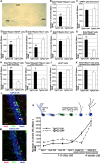
Figure 1. Activation of the PC3 Transgene Accelerates the Differentiation of 1–5-d-Old Adult-Generated Dentate Gyrus Stem and Progenitor Cells
(A) Selective targeting to the dentate gyrus in P95 mice of the PC3 transgene (X-gal staining, blue) conditionally activated since P30 by doxycycline treatment (scale bar, 615 μm). New stem and progenitor cells (B–F, J) as well as new post-mitotic neurons (G–I) 1–5-d-old were detected by incorporation of BrdU after five daily injections in P95 TgPC3 OFF and ON mice (the latter having the transgene active), and were classified by analyzing the expression of specific markers through multiple-labeling and confocal microscopy detection. (B) The number of putative stem cells (type-1; BrdU/GFAP/nestin-positive) decreased about 50% in mice with activated PC3 transgene. (C) This decrease was specific, given that the total number of GFAP-positive cells was only slightly reduced. (D) Activation of the PC3 transgene also reduced a the number of new progenitor cells type-2ab (BrdU/GFAP-positive and nestin-negative), (E) type-2b (BrdU/nestin/DCX-positive), and (F) type-3 (BrdU/DCX-positive and nestin-negative). (G) Conversely, the new differentiating early post-mitotic neurons at stage 5 and 6 (BrdU/NeuN-positive), or (H) those specifically at stage 6 (BrdU/DCX/NeuN-positive), presented a strong, significant increase in mice with activated PC3 transgene. (I) The new differentiating neurons, whose number is increased by activation of the PC3 transgene, also expressed NeuroD1, which is required for their differentiation [47]. (J) Nonetheless, in mice with active PC3 transgene, no significant change was observed in the total number of new 1–5-d-old neurons in dentate gyrus—as detected by BrdU incorporation—or (K) in the total number of new terminally differentiated neurons (4 wk old, positive to BrdU and NeuN). These findings indicate, as a whole, that activation of the PC3 transgene selectively accelerates the transition of stem and progenitor cells toward a differentiated state. The cell numbers shown in (B–K) were measured as described in Materials and Methods and are represented as mean ± SEM from the analysis of at least three animals per group. (L and M) Representative confocal images of new 1–5-d-old dentate gyrus neurons labeled by BrdU/DCX/NeuN-positive (stage 6; red, green, blue, respectively) in TgPC3 ON and OFF mice. The merge shows neurons positive for all three antigens as yellow cells present in TgPC3 ON, indicated by white arrowheads; scale bar, 25 μm. DG, dentate gyrus. *p < 0.05 versus TgPC3 OFF; **p < 0.02 versus TgPC3 OFF; Student's t-test. (N) Summary from data of Figure 1 of the effect of the activation of TgPC3 on new progenitor cells and post-mitotic neurons.
Figure 2
Figure 2. Morphological Analysis of the Development of Adult-Generated Dentate Gyrus Neurons
(A) Representative morphology of adult-generated neurons labeled with GFP by retrovirus-mediated gene transduction, analyzed from 7–28 dpi, in control (TgPC3 OFF) or activated transgenic mice (TgPC3 ON; the transgene was activated 60 d before infection, according to the time schedule of Figure 1). The GFP+ neurons (green) are localized at the hilar border of the dentate gyrus, whose cells are identified by Hoechst 33258 staining (blue). The molecular layer is localized at the upper side of the images. Scale bar, 50 μm. (B) Quantification of the dendritic length of adult-generated GFP+ neurons at 7, 16, and 28 dpi. (F(5, 148) = 73.7; p < 0.0001, ANOVA. *p < 0.05 or **p < 0.001 between TgPC3 ON and OFF groups at the same day post infection, ANOVA Fisher's PLSD post-hoc analysis). (C) Quantification of branching points at 7, 16, and 28 dpi. (F(5, 148) = 19.8; p < 0.0001, ANOVA. *p < 0.05 between 7 dpi TgPC3 ON and OFF groups, ANOVA Fisher's PLSD post-hoc analysis). (D) Quantification of spine density in the dendritic processes of GFP+ neurons analyzed at 28 dpi and 70 dpi. *p < 0.002, Student's t-test. Data in (B–D) are presented as mean ± SEM; at least three animals per group were analyzed. (E) Representative confocal images of dendrites and spines in 28 dpi and 70 dpi GFP+ dentate gyrus neurons. Scale bar, 12 μm.
Figure 3
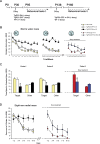
Figure 3. PC3 Transgene Activation Impairs Learning in the Morris Water Maze and Radial Maze
(A) Experimental timeline as a function of mouse age in days. P95 mice were subjected to a first session of spatial learning and memory tests (behavioral tests 1; TgPC3 ON mice had the transgene active starting from P30 throughout the experiment). At the end of the first session, TgPC3 OFF mice were treated with doxycycline to activate the PC3 transgene and subjected to a further experimental session (behavioral tests 2). Arrows indicate the starting days of doxycycline treatment. (B) TgPC3 ON (n = 10), TgPC3 OFF (n = 8) and WT (n = 23) mice were trained in the Morris water maze for 18 trials (six trials per day; trial/block 1–18), followed by 2 d of reversal learning (trial/block 19–30) in which the platform was moved to the opposite position. After the PC3 transgene activation, TgPC3 OFF (labelled as TgPC3 OFF → ON, n = 8) and WT-doxy (n = 11) mice were subjected to a new learning session (trial/block 31–42) in which the platform was moved to a novel position in the pool. Escape latency is expressed in seconds required to reach the platform. The performance of TgPC3 ON mice was significantly impaired during both learning (left) and reversal learning (middle) in comparison with both TgPC3 OFF and WT. TgPC3 OFF → ON mice were significantly impaired in the new learning (right) in comparison with WT-doxy. (C) Probe trials expressed as time (s) spent in the target quadrant (Target) or in the control quadrants (Other). Compared to TgPC3 OFF and WT, TgPC3 ON mice spent a significantly smaller amount of time in the target quadrant both in probe 1 (left) and probe 2 (middle). During probe 3 (right), TgPC3 OFF → ON mice spent a significantly smaller amount of time in the target quadrant compared to WT-doxy. (D) Mice were trained for 10 d in a fully baited, eight-arm radial maze. The learning performance is expressed as percentage of errors until eight correct arm choices have been observed or after the maximal permitted number of trials. The performance of TgPC3 ON mice (n = 10) was significantly impaired in comparison with those of TgPC3 OFF (n = 8) and WT (n = 23) (left). In session 2 of the behavioral tests, the performance of TgPC3 OFF → ON mice (n = 8) was significantly impaired in comparison with that of WT-doxy (n = 11) (right). *p < 0.05 in comparison with all other groups.
Figure 4
Figure 4. PC3 Transgene Activation Impairs Contextual but Not Cued Fear Conditioning
(A) Percentage of time spent in freezing behavior by TgPC3 ON (n = 8), TgPC3 OFF (n = 8), and WT (n = 21) mice during training in the conditioning box. Training consisted of an acclimatizing period (Pre-CS, 120 s) followed by a tone presentation (CS, 30 s) paired, in the last 2 s, with a foot-shock (US). After CS-US pairing, the animals were left in the conditioning box for a further 30 s (Post-US). During training, no significant differences in the level of freezing were observed among the different groups of mice. (B) Percentage of time spent in freezing behavior in the contextual test, carried out 24 h after training, by TgPC3 ON, TgPC3 OFF, and WT mice. TgPC3 ON mice showed a significant reduction in the level of freezing compared with TgPC3 OFF and WT (left). In session 2 of the behavioral tests, TgPC3 OFF → ON mice showed a significant memory impairment compared with WT-doxy (right). (C) Percentage of time spent in freezing behavior in the cued test, carried out 2 h after the contextual test in a different setting, by TgPC3 ON, TgPC3 OFF, and WT mice. No significant differences in the retention level were observed among the different groups of mice during either Pre-CS or CS phases (left). In session 2 of the behavioral tests, no significant differences in the level of freezing were observed between TgPC3 OFF → ON and WT-doxy mice (right). *p < 0.05 in comparison with all other groups.
Figure 5
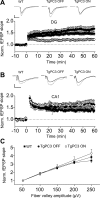
Figure 5. Effect of Premature Differentiation of Adult-Generated Granule Cells on Hippocampal Synaptic Plasticity
(A) LTP of lateral perforant pathway synapses in the dentate gyrus is reduced in TgPC3 ON but not in TgPC3 OFF mice. Upper panel shows traces of fEPSPs immediately before (black traces) and after 50 min (gray traces) of high-frequency trains in WT, TgPC3 ON, and OFF mice. Averages of 10 traces for each example are shown. The stimulus artifact was digitally removed for display purposes. Scales: 5 ms and 100 μV wt; 25 μV TgPC3 OFF; 60 μV TgPC3 ON. (B) LTP of Schaffer collateral synapses measured in CA1 was unaffected by early PC3 gene activation (p > 0.2). Upper panel: representative fEPSPs before (black traces) and after (gray traces) high frequency stimulations in all conditions. Shown are averages of 10 traces each. The stimulus artifact was digitally removed for display purposes. Scales: 5 ms and 60 μV wt; 100 μV TgPC3 OFF; 100 μV TgPC3 ON. (C) The slope of input-output (I/O) curves at perforant-path synapses in dentate gyrus was similar in all three conditions.
Figure 6
Figure 6. Lack of Integration into Dentate Gyrus Memory Circuits of New Neurons Derived from Dentate Gyrus Progenitor Cells Whose Differentiation Was Anticipated
New neurons derived from dentate gyrus progenitor cells whose differentiation was anticipated by the PC3 transgene do not become integrated into dentate gyrus spatial learning circuits. (A) Experimental design. 2, 4, and 6 wk after treatment with BrdU, TgPC3 ON/OFF and WT mice underwent Morris water maze behavioral training, which is expected to activate new neurons. (B) Representative confocal images of c-fos+ (blue), Brdu+ (red), NeuN+ (green; merged with c-fos+ and Brdu+) and of c-fos+/BrdU+/NeuN+ cells (white arrowhead) in the dentate gyrus following behavioral training in the 6-wk groups of TgPC3 ON and TgPC3 OFF mice (scale bar, 58 μm). (C) New activated neurons, identified by positivity to c-fos (c-fos+/BrdU+/NeuN+ cells), were quantified as percentage ratio to the number of new neurons (Brdu+/NeuN+). The fraction of new neurons activated by behavioral training was null in TgPC3 OFF mice undergoing behavioral training 2 weeks after treatment with BrdU (n = 3) but became significantly detectable 4 and 6 wk after BrdU treatment (n = 6 at both time points; F(5, 274) = 7.4; p < 0.0001, ANOVA. *p < 0.05 or **p < 0.02 versus the other groups, ANOVA Fisher's PLSD post-hoc analysis). By contrast, no activation of new neurons was observed in the 2-, 4-, and 6-wk groups of TgPC3 ON mice (n = 3, 6, and 6, respectively). (D) New neurons were generated with equivalent frequency in the TgPC3 ON and TgPC3 OFF groups, as indicated by the similar percentage ratio between number of new neurons (BrdU+/NeuN+) and total number of neurons (NeuN+ cells) identified 2, 4, and 6 wk after BrdU treatment (F(5, 122) = 0.24; p > 0.05, ANOVA). (E) After behavioral training, the presence of activated neurons in the whole neuronal population of the dentate gyrus—measured as percentage ratio of c-fos+/NeuN+ to NeuN+ cells—was similar in all groups of TgPC3 OFF mice, whereas in TgPC3 ON mice the presence of activated neurons was significantly reduced in the 4- and 6-wk groups, indicating a progressive decrease in the population of active neurons (F(5, 112) = 16.0; p < 0.0001, ANOVA. *p < 0.02 versus 2-, 4-, 6-wk TgPC3 OFF and 2-wk TgPC3 ON mice groups, ANOVA Fisher's PLSD post-hoc analysis). The percentage ratios shown in (C–E) were calculated from absolute cell numbers measured as described in Materials and Methods and are represented as mean ± SEM.

Similar articles

Cited by

References

    1. Altman J, Das GD. Autoradiographic and histological evidence of post-natal hippocampal neurogenesis in rats. J Comp Neurol. 1965;124:319–335. - PubMed
    1. Gould E, Reeves AJ, Fallah M, Tanapat P, Gross CG, et al. Hippocampal neurogenesis in adult Old World primates. Proc Natl Acad Sci U S A. 1999;96:5263–5267. - PMC - PubMed
    1. Nottebohm F. Neuronal replacement in adult brain. Brain Res Bull. 2002;57:737–749. - PubMed
    1. Bayer SA, Yackel JW, Puri PS. Neurons in the rat dentate gyrus granular layer substantially increase during juvenile and adult life. Science. 1982;216:890–892. - PubMed
    1. Eriksson PS, Perfilieva E, Bjork-Eriksson T, Alborn AM, Nordborg C, et al. Neurogenesis in the adult human hippocampus. Nat Med. 1998;4:1313–1317. - PubMed

Publication types

MeSH terms