Review
. 2009:71:59-82.
doi: 10.1146/annurev.physiol.010908.163204.

Plant ion channels: gene families, physiology, and functional genomics analyses

Affiliations
Review

Plant ion channels: gene families, physiology, and functional genomics analyses

John M Ward et al. Annu Rev Physiol. 2009.

Abstract

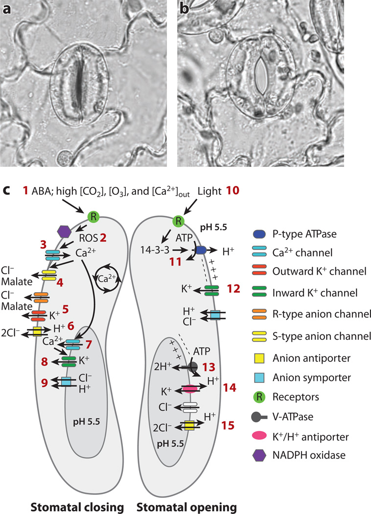
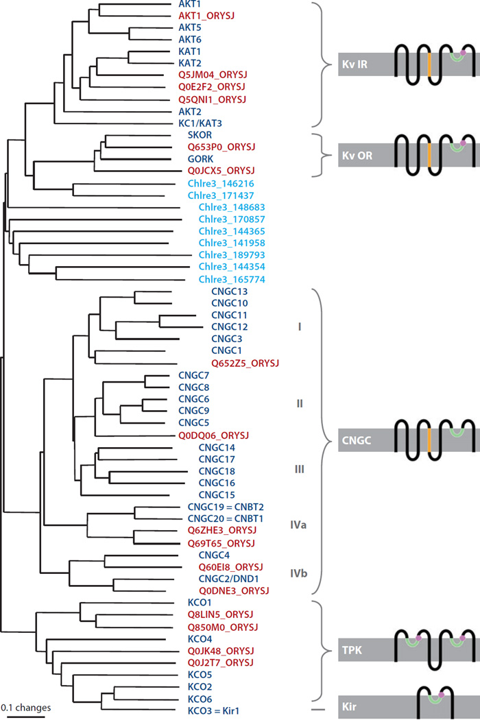
Distinct potassium, anion, and calcium channels in the plasma membrane and vacuolar membrane of plant cells have been identified and characterized by patch clamping. Primarily owing to advances in Arabidopsis genetics and genomics, and yeast functional complementation, many of the corresponding genes have been identified. Recent advances in our understanding of ion channel genes that mediate signal transduction and ion transport are discussed here. Some plant ion channels, for example, ALMT and SLAC anion channel subunits, are unique. The majority of plant ion channel families exhibit homology to animal genes; such families include both hyperpolarization- and depolarization-activated Shaker-type potassium channels, CLC chloride transporters/channels, cyclic nucleotide-gated channels, and ionotropic glutamate receptor homologs. These plant ion channels offer unique opportunities to analyze the structural mechanisms and functions of ion channels. Here we review gene families of selected plant ion channel classes and discuss unique structure-function aspects and their physiological roles in plant cell signaling and transport.


Figures

Figure 1
Figure 1
Model for ion channel and transporter classes and their demonstrated or predicted roles during stomatal movements. (a) A closed stomate in a leaf from Vicia faba (broad bean). (b) An open stomate from Vicia faba. Two guard cells surround the stomatal pore and regulate the aperture of the central pore. (c) Model for regulation and activity of guard cell ion channels and transporters. (Left) (1) Signals that induce stomatal closing include the hormone abscisic acid (ABA), high [CO2], high extracellular [Ca2+], and high ozone [O3]. Many of the receptors involved remain to be identified (see Reference 26). Stomatal closing requires net cellular efflux of solutes, in particular K+, Cl, and malate. ABA induces reactive oxygen species (ROS) production (2), which activates Ca2+-permeable ICa channels (3). Cytosolic Ca2+ concentration ([Ca2+]cyt) is a central regulator of transport mechanisms in guard cells and activates slow/sustained (S-type) anion channels (4) and vacuolar SV (TPC) channels (7) and VK (TPK) channels (8). Cl and malate efflux through S-type and rapid (R-type) anion channels (4) and possibly CLC Cl/H+ antiporters (6) causes depolarization and drives K+ efflux through outward-rectifying K+ channels (5). At the vacuole membrane, SV (TPC1) channels (7) are Ca2+-activated and Ca2+-permeable voltage-dependent channels. VK (TPK) channels (8) are Ca2+ activated, are highly selective for K+, and are proposed to allow K+ release from the vacuole during stomatal closing. (Right) Mechanisms that function in guard cell ion uptake and stomatal opening. Blue light (10) activates phototropins (receptor/kinase), leading to stomatal opening. Signaling results in 14-3-3 binding to plasma membrane proton pumps (11), leading to hyperpolarization and acidification of the extracellular space. Hyperpolarization activates inward-rectifying K+ channels (12). At the vacuolar membrane, proton pumps (13) acidify the vacuole lumen and drive K+/H+ antiporters (14 ). Cl and malate may accumulate in the vacuole through anion channel and anion antiporters (15 ).
Figure 2
Figure 2
Phylogenetic tree of P-loop-containing proteins from Arabidopsis thaliana (dark blue), rice (Oryza sativa; red ), and Chlamydomonas reinhardtii (light blue). Where applicable, schematized membrane topologies are indicated: P-loop, green; transmembrane domain 4 (TM4) including positively charged amino acids, orange; K+ selectivity filter residues, magenta. Cytosolic elements such as long C-terminal domains, cyclic nucleotide (cNMP) binding domains, or ankyrin repeats are not depicted. The photosynthetic alga C. reinhardtii has a unique set of ion channel genes (see Reference 105 and text). The P-loop-containing channels from C. reinhardtii all have a positively charged TM4, but only five (179857, 144365, 189793, 144354, and 165774) have a K+ selectivity filter. Because these do not form a clade, and because the C. reinhardtii channels are poorly resolved in general, they could not be attributed to specific classes. Scale indicates the number of amino acid substitutions per site. Kv IR, inward-rectifying K+ channel; Kv OR, outward-rectifying K+ channel; CNGC, cyclic nucleotide–gated channel; TPK, two-pore K+ channel.

References

    1. Cole KS, Curtis HJ. Electrical impedance of Nitella during activity. J. Gen. Physiol. 1938;22:37–64. - PMC - PubMed
    1. Hodgkin AL, Huxley AF. The components of membrane conductance in the giant axon of Loligo. J. Physiol. 1952;116:473–496. - PMC - PubMed
    1. Gaffey CT, Mullins LJ. Ion fluxes during the action potential in Chara . J. Physiol. 1958;144:505–524. - PMC - PubMed
    1. Hope AB, Findlay GP. The action potential in Chara. Plant Cell Physiol. 1962;5:377–379.
    1. Hodick D, Sievers A. The action potential of Dionaea muscipula Ellis. Planta. 1988;174:8–18. - PubMed

Publication types

LinkOut - more resources