Review
. 2009:4:287-313.
doi: 10.1146/annurev.pathol.4.110807.092246.

Ovarian cancer

Affiliations
Review

Ovarian cancer

Kathleen R Cho et al. Annu Rev Pathol. 2009.

Abstract

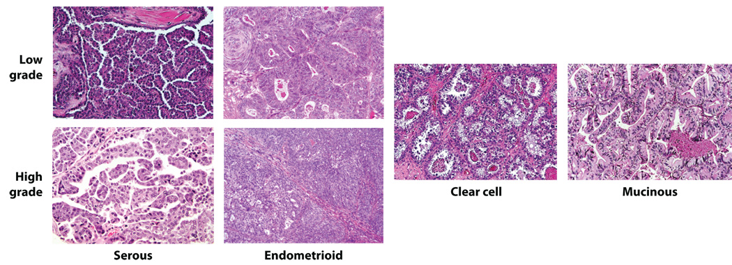
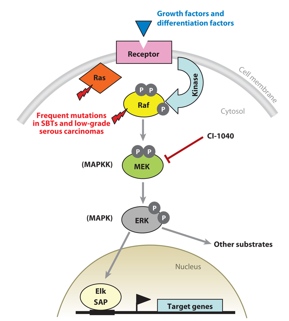
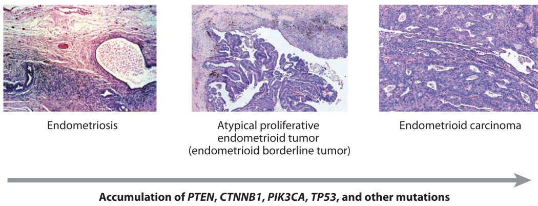
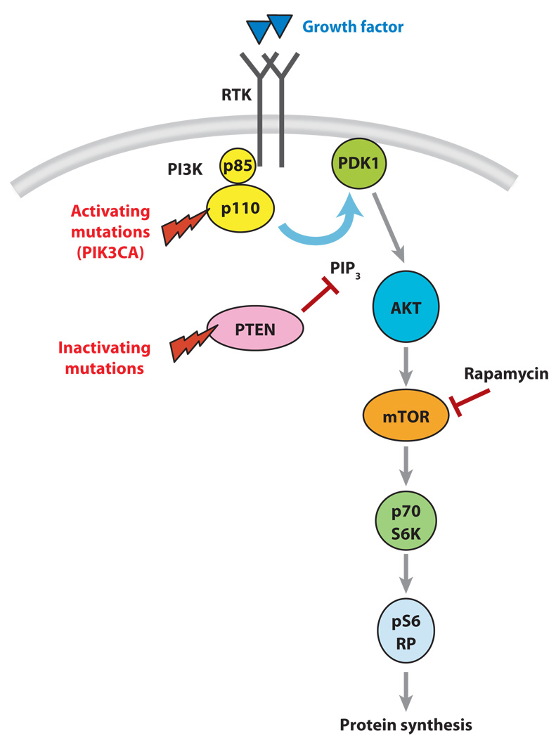
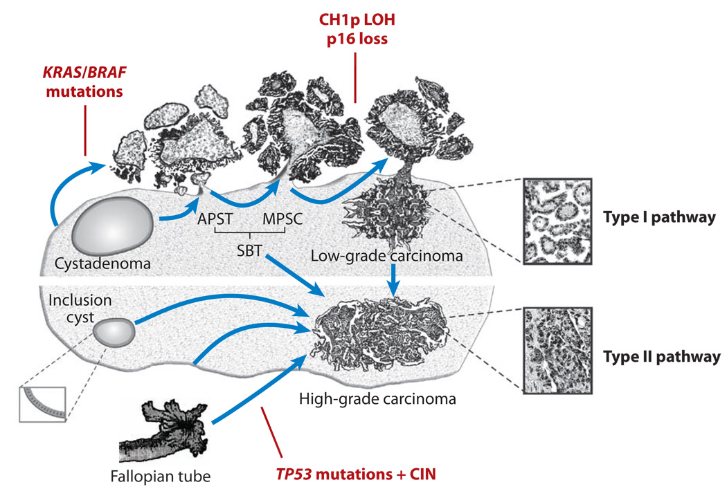
Ovarian carcinomas are a heterogeneous group of neoplasms and are traditionally subclassified based on type and degree of differentiation. Although current clinical management of ovarian carcinoma largely fails to take this heterogeneity into account, it is becoming evident that each major histological type has characteristic genetic defects that deregulate specific signaling pathways in the tumor cells. Moreover, within the most common histological types, the molecular pathogenesis of low-grade versus high-grade tumors appears to be largely distinct. Mouse models of ovarian carcinoma have been developed that recapitulate many of the morphological features, biological behavior, and gene-expression patterns of selected subtypes of ovarian cancer. Such models will likely prove useful for studying ovarian cancer biology and for preclinical testing of molecularly targeted therapeutics, which may ultimately lead to better clinical outcomes for women with ovarian cancer.


Figures

Figure 1
Figure 1. Representative examples of the major histological types of ovarian carcinoma
Figure 2
Figure 2. Schematic illustration of the RAS-RAF-MEK-ERK (MAPK) signaling pathway
This cell signaling pathway is important for the cellular response to a variety of growth and differentiation factors. Aberration of this pathway in ovarian SBTs and low-grade serous carcinomas is mainly due to activating mutations of KRAS and BRAF, which result in constitutive activation of MAPK-mediated signaling in these tumors. Activated MAPK signaling alters expression of downstream target genes, including up-regulation of cyclin D1. Several MEK small compound inhibitors such as CI-1040 have been generated. These inhibitors effectively suppress MAPK activation and hold promise for treatment of patients with advanced stage low-grade serous carcinomas.
Figure 3
Figure 3. Histological and molecular genetic progression of ovarian endometrioid carcinoma
Low-grade endometrioid carcinomas often arise from endometrioid borderline tumors, which in turn may arise from endometriosis. This step-wise histopathological progression is often accompanied by accumulation of mutations predicted to deregulate canonical Wnt signaling (usually CTNNB1) and/or PI3K/Pten signaling (PTEN, PIK3CA). TP53 mutations are usually observed in high-grade endometrioid carcinomas in the absence of Wnt and PI3K/Pten pathway defects.
Figure 4
Figure 4. Schematic illustration of one aspect of PI3K/Pten signaling
Activating mutations of PIK3CA or inactivating mutations of PTEN result in activation of Akt-mediated signaling to mTOR and other downstream effectors that affect protein synthesis. Rapamycin is a well known inhibitor of mTOR. GF, growth factor; RTK, receptor tyrosine kinase.
Figure 5
Figure 5. Genetically engineered mouse models of ovarian cancer recapitulate their human counterparts and respond to molecularly-targeted therapeutics
A) Hematoxylin & eosin stained section of endometrioid carcinoma-like mouse tumor. B) Representative ovarian carcinoma in right ovary of Apcflox/flox;Ptenflox/flox mouse 11 wks after ovarian bursal injection of adenovirus expressing Cre recombinase (AdCre). C) Rapamycin inhibits tumor growth. Animals with small ovarian carcinomas (6 wks after AdCre injection) were treated twice weekly for 4 wks with vehicle or two different doses of rapamycin. D) Representative ovarian carcinoma in right ovary after 4 wks of treatment with rapamycin.
Figure 6
Figure 6. Schematic illustration of Type I and Type II ovarian serous carcinoma pathogenesis
Development of ovarian high-grade and low-grade serous carcinomas involves two distinct pathways. Low-grade (Type I) carcinomas arise from serous borderline tumors (SBTs), which in turn develop from serous cystadenomas. This stepwise tumor progression in the low-grade pathway contrasts with the rapid progression pathway of high-grade (Type II) carcinomas, for which precursor lesions are not well recognized. High-grade carcinomas may arise from ovarian surface inclusions, peritoneal mesothelium or the distal portion (fimbriae) of the fallopian tube. These carcinomas disseminate to pelvic and peritoneal organs early in their progression.

References

    1. Jemal A, Siegel R, Ward E, Murray T, Xu J, Thun MJ. Cancer statistics, 2007. CA Cancer J Clin. 2007;57:43–66. - PubMed
    1. Feeley KM, Wells M. Precursor lesions of ovarian epithelial malignancy. Histopathology. 2001;38:87–95. - PubMed
    1. Bell DA. Origins and molecular pathology of ovarian cancer. Mod Pathol. 2005;18 Suppl 2:S19–S32. - PubMed
    1. Dubeau L. The cell of origin of ovarian epithelial tumors and the ovarian surface epithelium dogma: does the emperor have no clothes? Gynecol Oncol. 1999;72:437–442. - PubMed
    1. Kurman RJ. Blaustein's pathology of the female genital tract. 5th ed. New York: Springer-Verlag; 2002.

Publication types