

Review
doi: 10.1098/rsbl.2008.0439.
Telencephalon enlargement by the convergent evolution of expanded subventricular zones
Affiliations
- PMID: 18842571
- PMCID: PMC2657736
- DOI: 10.1098/rsbl.2008.0439
Item in Clipboard
Review
Telencephalon enlargement by the convergent evolution of expanded subventricular zones
Biol Lett.
.
Abstract
Some mammals and birds independently evolved an enlarged telencephalon. They appear to have done so, at least in part, by developing a thick telencephalic subventricular zone (SVZ). We suggest that this correlation between telencephalic enlargement and SVZ expansion is due to a mechanical constraint acting on the proliferative ventricular zone (VZ). Essentially, we argue that rapid proliferation in the VZ after post-mitotic cells in the overlying mantle zone have begun to form limits the VZ's tangential expandability and forces some proliferating cells to emigrate from the VZ and expand the pool of proliferating cells that comprise the SVZ.
Figures
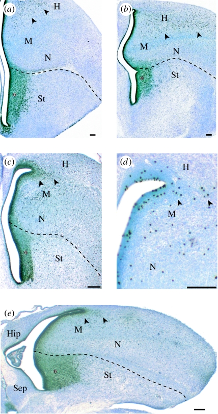
Sections through the telencephalon of (a, b) parakeets and (c–e) zebra finches labelled with antibodies against (a–c, e) proliferating cells (anti-PCNA) or (d) mitotic cells (anti-phosphorylated histone H3). The thick SVZ in the striatum (St) is marked with an asterisk. Mitotic cells in the hyperpallium, which is probably homologous to at least part of the mammalian neocortex, are indicated with arrowheads. H, hyperpallium; M, mesopallium; N, nidopallium; Hip, hippocampus; Sep, septum. Scale bar, 100 μm.
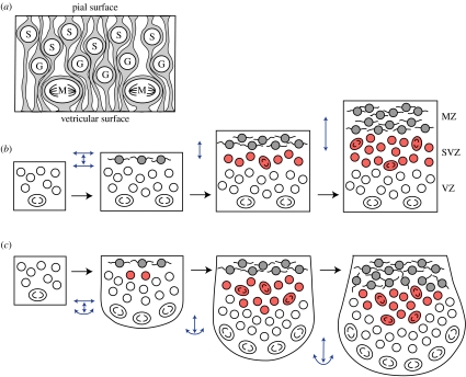
(a) In the VZ, most mitoses (M) occur at the ventricular surface; G- and S-phase cells are located away from the ventricle. (b) Early in embryogenesis, the VZ expands mainly tangentially. After the MZ has formed, tangential VZ expansion is limited by the processes of post-mitotic MZ cells. This, in turn, increases congestion within the VZ to the point where some proliferating cells emigrate from the VZ and form the SVZ. (c) If the mechanical coupling between the VZ and the MZ is low, then a rapidly proliferating VZ may continue to expand tangentially when the MZ does not. In such cases, an intraventricular ridge with a large SVZ is formed. Blue arrows indicate radial and tangential expansion along the pial and ventricular surfaces.
References
-
- Abdel-Mannan O., Cheung A.F., Molnár Z. Evolution of cortical neurogenesis. Brain Res. Bull. 2008;75:398–404. doi:10.1016/j.brainresbull.2007.10.047 - DOI - PubMed
-
- Boire D., Baron G. Allometric comparison of brain and main brain subdivisions in birds. J. Hirnforsch. 1994;35:49–66. - PubMed
-
- Charvet C.J., Striedter G.F. Developmental species differences in brain cell cycle rates between Northern bobwhite quail (Colinus virginianus) and parakeets (Melopsittacus undulatus): implications for mosaic brain evolution. Brain Behav. Evol. 2008;72:295–306. doi:10.1159/000184744 - DOI - PubMed
-
- Chenn A., Walsh C.A. Regulation of cerebral cortical size by control of cell cycle exit in neural precursors. Science. 2002;297:365–369. doi:10.1126/science.1074192 - DOI - PubMed
Publication types
MeSH terms
LinkOut - more resources
Full Text Sources