. 2008 Dec 12;283(50):35173-85.
doi: 10.1074/jbc.M803219200. Epub 2008 Oct 7.

Human Wrnip1 is localized in replication factories in a ubiquitin-binding zinc finger-dependent manner

Affiliations

Human Wrnip1 is localized in replication factories in a ubiquitin-binding zinc finger-dependent manner

Nicola Crosetto et al. J Biol Chem. .

Abstract

Wrnip1 (Werner helicase-interacting protein 1) has been implicated in the bypass of stalled replication forks in bakers' yeast. However, the function(s) of human Wrnip1 has remained elusive so far. Here we report that Wrnip1 is distributed inside heterogeneous structures detectable in nondamaged cells throughout the cell cycle. In an attempt to characterize these structures, we found that Wrnip1 resides in DNA replication factories. Upon treatments that stall replication forks, such as UVC light, the amount of chromatin-bound Wrnip1 and the number of foci significantly increase, further implicating Wrnip1 in DNA replication. Interestingly, the nuclear pattern of Wrnip1 appears to extend to a broader landscape, as it can be detected in promyelocytic leukemia nuclear bodies. The presence of Wrnip1 into these heterogeneous subnuclear structures requires its ubiquitin-binding zinc finger (UBZ) domain, which is able to interact with different ubiquitin (Ub) signals, including mono-Ub and chains linked via lysine 48 and 63. Moreover, the oligomerization of Wrnip1 mediated by its C terminus is also important for proper subnuclear localization. Our study is the first to reveal the composite and regulated topography of Wrnip1 in the human nucleus, highlighting its potential role in replication and other nuclear transactions.


Figures
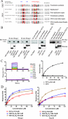
FIGURE 1.
FIGURE 1.
Wrnip1 is a mono- and poly-Ub-binding protein. A, Wrnip1 contains a UBZ4 domain. Multiple alignment of UBZ3 (upper part) and UBZ4 (lower part) zinc finger domains is shown. Invariant or conserved in at least 40% of the sequences residues are shown on a black or gray background, respectively. The two uppermost lines indicate the secondary structure (E = extended, H = helix) of polymerase η and the residues (asterisks) with NMR signals that were perturbed upon Ub binding (17). The beginning and the end of each domain are indicated by the number of their first and last amino acids, respectively. Accession numbers are shown for each protein and refer to the NCBI Protein data base. Known or proposed functions of each protein are indicated on the right. B, Wrnip1 UBZ is necessary for interaction with mono-Ub. Left panel, GST-Ub and Ub-agarose pulldown of mouse FLAG-Wrnip1 overexpressed in HEK 293T cells. 2nd panel from the left, GST-Ub I44A pulldown of M. musculus (Mm) FLAG-Wrnip1 overexpressed in HEK 293T. 2nd panel from the right, GST-Ub pulldown of M. musculus FLAG-Wrnip1 D37A overexpressed in HEK 293T. Right panel, pulldown of Myc-tagged human Rad18 wild-type and D221A with GST-Ub. IB, immunoblot. C, Wrnip1 UBZ is a mono-Ub-interacting domain. Left graphs, sensorgrams obtained for different concentrations of free mono-Ub flown over wild-type (upper left graph) and D37A (lower left graph) GST-UBZWrnip1. KD values were calculated by fitting a Langmuir binding isotherm to the data (right graph). D, Wrnip1 and Rad18 UBZs have a different selectivity for di-ubiquitin chains. Isolated UBZs of Wrnip1 (left graph) and Rad18 (right graph) were coupled to a chip for SPR, and responses to increasing concentrations of either mono-Ub, Lys-48-, or Lys-63-linked di-ubiquitin chains were measured. The equilibrium responses obtained were plotted against Ub concentration, and KD values were calculated from these Langmuir binding isotherms.
FIGURE 2.
FIGURE 2.
Wrnip1 is concentrated in various subnuclear structures. A, Wrnip1 is localized in punctated and patch-like structures in the absence of induced DNA damage. Endogenous Wrnip1 in HeLa and MRC5 cells (left panels). Endogenous and recombinant Wrnip1 fully co-localize in HeLa cells stably expressing low levels of Wrnip1-EGFP (right panels). Wrnip1-EGFP was detected using the fluorescence signal from EGFP, whereas endogenous Wrnip1 was probed with a commercially available anti-Wrnip1 antibody. B, some Wrnip1 foci correspond to replication factories. HeLa_Wrnip1-EGFP cells were stained with an antibody against RPA. Wrnip1-EGFP was detected using the fluorescence signal from EGFP. Rectangles on the top and right side of each squared image represent signal profiles along the z axis. Histograms on the right, quantification of the percentage of Wrnip1-EGFP foci overlapping with RPA and of RPA foci co-localizing with Wrnip1. C, some Wrnip1 foci overlap with PML bodies. HeLa_Wrnip1-EGFP cells were stained with an antibody against PML. In all the images, white arrows indicate examples of co-localizing proteins and blue arrows abutting foci.
FIGURE 3.
FIGURE 3.
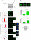
Wrnip1 foci are dynamic. A, Wrnip1 foci are visible throughout the cell cycle. HeLa_Wrnip1-EGFP cells were synchronized by a double thymidine block. Top, the overall amount of endogenous Wrnip1 remains constant throughout the cell cycle. WCE, whole cell extract. Left column panels, cell cycle phase distributions assessed by propidium iodide stain and cytofluorimetry. Right column panels, low magnification fields of asynchronized or synchronized HeLa cells. The small panels show representative magnifications of one or two cells found in the bigger adjacent panels. The five small panels boxed in red display nuclei in subsequent phases of mitosis. White arrows mark Wrnip1 signal at the cleavage furrow. Wrnip1-EGFP was detected using EGFP fluorescence. IB, immunoblot. DAPI, 4′,6-diamidino-2-phenylindole. B, Wrnip1 is re-distributed following DNA damage. Top, HeLa_Wrnip1-EGFP cells were treated with the indicated DNA-damaging agents, extracted, fixed, and used for microscopy. In the middle, quantification of the images above. Each green histogram represents mean values, whereas black bars mark standard deviations. P, result of a Student's t test comparing the indicated distributions. Bottom, the overall amount of Wrnip1 does not change upon UVC treatment. WCE, whole cell extract; IB, immunoblot; HU, hydroxyurea.
FIGURE 4.
FIGURE 4.
Wrnip1 UBZ is essential for its localization to nuclear foci. A, Wrnip1 and Rad18 UBZ are indispensable for their presence in nuclear foci, independently of DNA damage. Left panels, HeLa_Wrnip1-EGFP either wild-type or D37A as well as HeLa cells transfected with Wrnip1 ΔUBZ-EGFP were extracted, fixed, and used for microscopy. Right panels, HeLa_Wrnip1-EGFP cells transfected with either wild-type or D221A human Myc-tagged Rad18 were similarly processed. Wrnip1-EGFP was detected using EGFP fluorescence, Myc-Rad18 using an anti-Myc antibody. B, Wrnip1 and Rad18 co-localization is significantly enhanced by UVC irradiation. HeLa_Wrnip1-EGFP cells were transfected with Myc-tagged Rad18 and either treated with UVC irradiation (right) or not (left). The white square encloses the area that is magnified in the small panels. Wrnip1-EGFP was detected using the fluorescence from EGFP, whereas Myc-Rad18 using anti-Myc antibodies. Histograms on the right, quantification of Wrnip1-positive foci containing also Rad18 and of Rad18-positive foci containing Wrnip1, with or without prior UVC irradiation. The distributions were compared by a Student's t test, and the calculated p values are displayed. C, type 4 UBZ is required for Wrnip1 and Rad18 to be localized in foci. Left, HeLa cells transfected with Wrnip1-EGFP wild-type and various UBZ chimeras. Right, HeLa cells transfected with Myc-Rad18 wild-type and UBZWrnip1 chimera. Wrnip1-EGFP was detected using EGFP fluorescence, Myc-Rad18 using an anti-Myc antibody.
FIGURE 5.
FIGURE 5.
Wrnip1 oligomerization is UBZ-independent and contributes to its presence in foci. A, Ub-binding ability of Wrnip1 is not required for oligomerization. Yeast two-hybrid assay with the Y190 strain. Empty, control yeast transformed with the indicated empty vectors. D37A, yeast transformed with Wrnip1 D37A. 1-250, 200-400, 397-665, yeast transformed with fragments of Wrnip1 encompassing amino acids from 1 to 250; from 200 to 400, and from 397 to 665, respectively. B, Wrnip1 UBMPolι chimera oligomerizes with wild-type Wrnip1. HEK 293T cells were transfected with FLAG-Wrnip1 together with either Wrnip1-EGFP or Wrnip1 UBMPolι-EGFP chimera. Protein complexes were immune precipitated with an anti-FLAG antibody and visualized by SDS-PAGE and Western blot. *, mono-ubiquitylated Wrnip1. IB, immunoblot. C, Wrnip1 UBZ is a monomer in solution. MALLS using purified UBZWrnip1 cleaved from GST by thrombin. The left peak represents Wrnip1 UBZ, and the right peak is from small molecules. Calculated and measured molecular weights of UBZWrnip1 are shown. D, C terminus of Wrnip1 is required for its presence in foci. HeLa_Wrnip1-EGFP cells were transfected with FLAG-tagged Wrnip1 wild type or lacking the last 215 amino acids.

References

    1. Branzei, D., and Foiani, M. (2008) Nat. Rev. Mol. Cell Biol. 9297 -308 - PubMed
    1. Moldovan, G. L., Pfander, B., and Jentsch, S. (2007) Cell 129665 -679 - PubMed
    1. Hishida, T., Iwasaki, H., Ohno, T., Morishita, T., and Shinagawa, H. (2001) Proc. Natl. Acad. Sci. U. S. A. 988283 -8289 - PMC - PubMed
    1. Hickson, I. D. (2003) Nat. Rev. Cancer 3169 -178 - PubMed
    1. Kudlow, B. A., Kennedy, B. K., and Monnat, R. J., Jr. (2007) Nat. Rev. Mol. Cell Biol. 8 394-404 - PubMed

Publication types

MeSH terms