. 2008 Nov 28;283(48):33329-36.
doi: 10.1074/jbc.M803726200. Epub 2008 Oct 8.

APOBEC3G subunits self-associate via the C-terminal deaminase domain

Affiliations

APOBEC3G subunits self-associate via the C-terminal deaminase domain

Ryan P Bennett et al. J Biol Chem. .

Abstract

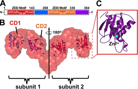
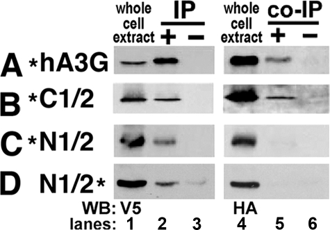
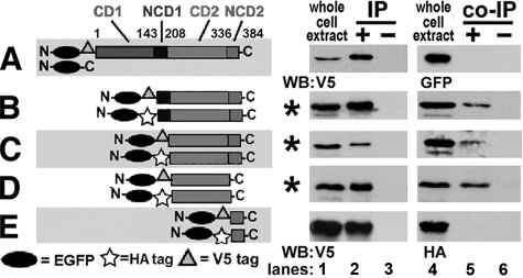
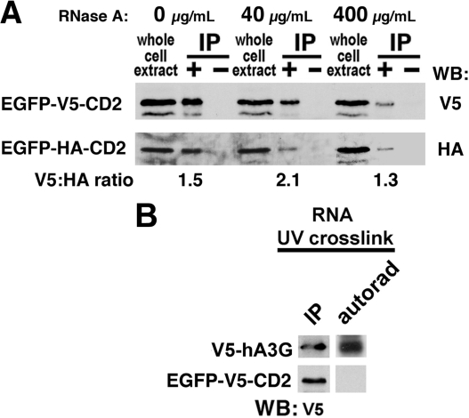
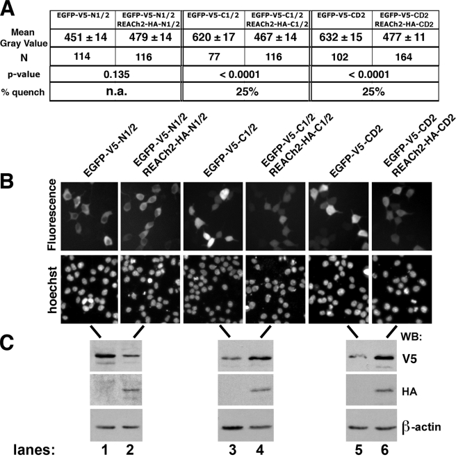
Human APOBEC3G (hA3G) is a cytidine deaminase active on HIV single-stranded DNA. Small angle x-ray scattering and molecular envelope restorations predicted a C-terminal dimeric model for RNA-depleted hA3G in solution. Each subunit was elongated, suggesting that individual domains of hA3G are solvent-exposed and therefore may interact with other macromolecules even as isolated substructures. In this study, co-immunoprecipitation and in-cell quenched fluorescence resonance energy transfer assays reveal that hA3G forms RNA-independent oligomers through interactions within its C terminus. Residues 209-336 were necessary and sufficient for homoligomerization. N-terminal domains of hA3G were unable to multimerize but remained functional for Gag and viral infectivity factor (Vif) interactions when expressed apart from the C terminus. These findings corroborate the small angle x-ray scattering structural model and are instructive for development of high throughput screens that target specific domains and their functions to identify HIV/AIDS therapeutics.


Figures

FIGURE 1.
FIGURE 1.
Schematic diagram of hA3G domain boundaries. A, hA3G primary structure showing residues of CD1 (red), NCD1 (blue), CD2 (orange), and NCD2 (violet) with the consensus ZDD motif shown within CD1 and CD2. N, N terminus; C, C terminus. B, the molecular envelope of the tail-to-tail dimer of hA3G based on SAXS analysis with docked CD domains indicated within the model. C, the relation between the hA3G volumes and the minimal catalytic domain from a related crystal structure. Dashed red boxes (B) show how volumes in the SAXS envelope accommodate a representative CD domain located by computational analysis in our previous publication (35).
FIGURE 2.
FIGURE 2.
Co-IPs of alternatively tagged hA3G and hA3G halves. A–D, the hA3G constructs co-transfected into 293T cells are represented in the left column with a * indicating the location of the tags (N- or C-terminal HA or V5) for each construct. Lanes 1 and 4 are blots showing the expression of each transfected protein in whole cell extracts. Lanes 2 and 5 are blots showing immunoprecipitated and co-immunoprecipitated proteins; all immunoprecipitations were performed with V5 antibody. Lanes 3 and 6 are controls for nonspecific binding to protein A beads. The antibody used for Western blotting (WB) is indicated on the bottom left for Western blots.
FIGURE 3.
FIGURE 3.
Self-association of C-terminal domains of hA3G. In A–E, the hA3G domains co-transfected into 293T cells are represented in the left column as horizontal gray bars for CD1, NCD1, CD2, and NCD2 with symbols for EGFP, HA, and V5 tags. On the right, lanes are depicted as in Fig. 2. A, negative control for EGFP and EGFP-V5-hA3G. N, N terminus; C, C terminus. B–E, co-IPs of alternatively tagged hA3G domains with conditions in which co-immunoprecipitation was successful indicated by *. WB, Western blotting.
FIGURE 4.
FIGURE 4.
Evidence for protein-protein self-association within CD2. A, co-immunoprecipitations of CD2 domain with increasing amounts of RNase A (0, 40, and 400 μg/ml). The ratio of V5 and HA Western signals from the same exposure for each condition is indicated below each immunoprecipitation lane with densitometry of each band determined using ImageJ software (National Institutes of Health). WB, Western blotting. B, 32P-radiolabeled RNA was analyzed for UV cross-linking to V5-hA3G or EGFP-V5-CD2 in cell extracts followed by RNase digestion and immunoprecipitation, as described under “Experimental Procedures.” On the left are V5 blots detecting the full-length hA3G and the CD2 domain after immunoprecipitation. On the right, the autorad lane reveals the presence or absence of radioactivity associated with the matched Western band. The results are indicative of low or no RNA binding for CD2 relative to intact hA3G.
FIGURE 5.
FIGURE 5.
In vivo FqRET of hA3G domains. A, as a measure of fluorescence intensity, the gray values from individual cells (number (N) indicated) in three separate fields were determined using ImageJ software (National Institutes of Health). The mean gray value is shown ± the S.E. The p values were determined by a linear regression model used to specify the relationship between the measure of fluorescence intensity in the presence and absence of REACh2-tagged domains as described under “Experimental Procedures.” The percentage of quench was measured as the difference in mean gray values of EGFP-tagged domains without and with REACh2, divided by the mean gray value for EGFP-tagged domains when expressed alone for C1/2 and CD2 domains. A quench value was not applicable (n.a.) for N1/2 because its p value revealed no significant difference (p > 0.05) with and without REACh2, suggesting that no quenching occurred. B, representative fields from images used to calculate fluorescence in A. EGFP fluorescence is shown above with Hoechst staining of the same field shown below representing live cell nuclei. C, Westerns with V5 and HA antibodies show relative tagged protein abundance for the transfected constructs, and the Western blot (WB) with β-actin antibody shows an equivalent load of cell extract for each condition. The transfected constructs indicated above B are the same for the lanes in B and C.

References

    1. MacElrevey, C., and Wedekind, J. E. (2008) in RNA and DNA Editing: Molecular Mechanisms and Their Integration into Biological Systems (Smith, H. C., ed) pp. 369-419, Wiley, New York
    1. Wedekind, J. E., Dance, G. S., Sowden, M. P., and Smith, H. C. (2003) Trends Genet. 19 207-216 - PubMed
    1. Jarmuz, A., Chester, A., Bayliss, J., Gisbourne, J., Dunham, I., Scott, J., and Navaratnam, N. (2002) Genomics 79 285-296 - PubMed
    1. Sheehy, A., Gaddis, N., Choi, J., and Malim, M. (2002) Nature 418 646-650 - PubMed
    1. Mehle, A., Wilson, H., Zhang, C., Brazier, A. J., McPike, M., Pery, E., and Gabuzda, D. (2007) J. Virol. 81 13235-13241 - PMC - PubMed

Publication types

MeSH terms

Substances

LinkOut - more resources