Case Reports
. 2009 Jan;30(1):113-9.
doi: 10.3174/ajnr.A1305. Epub 2008 Oct 8.

Pontine tegmental cap dysplasia: MR imaging and diffusion tensor imaging features of impaired axonal navigation

Affiliations
Case Reports

Pontine tegmental cap dysplasia: MR imaging and diffusion tensor imaging features of impaired axonal navigation

P Jissendi-Tchofo et al. AJNR Am J Neuroradiol. 2009 Jan.

Abstract

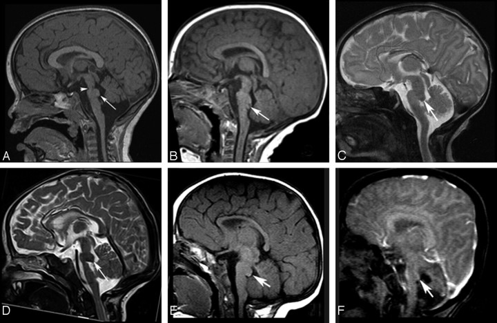
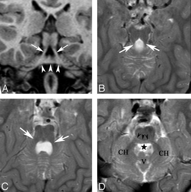
Background and purpose: Malformations of the brain stem are uncommon. We present MR imaging and diffusion tensor imaging (DTI) features of 6 patients with pontine tegmental cap dysplasia, characterized by ventral pontine hypoplasia and a dorsal "bump," and speculate on potential mechanisms by which it forms.

Materials and methods: Birth and developmental records of 6 patients were reviewed. We reviewed MR imaging studies of all patients and DTIs of patient 3. Potential developmental causes were evaluated.

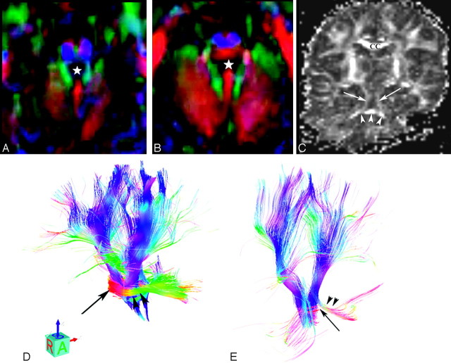
Results: All patients were born uneventfully after normal pregnancies except patient 6 (in utero growth retardation). They presented with multiple cranial neuropathies and evidence of cerebellar dysfunction. Variable hypotonia and motor dysfunction were present. Imaging revealed ventral pontine hypoplasia and mild cerebellar vermian hypoplasia, in addition to an unusual rounded to beaklike "bump" on the dorsal surface of the pons, extending into the fourth ventricle. Color fractional anisotropy maps showed the bump to consist of a bundle of axons directed horizontally (left-right). The bump appeared, on morphologic images, to be continuous with the middle cerebellar peduncles (MCPs), which were slightly diminished in size compared with those in healthy infants. Analysis of the DTI was, however, inconclusive regarding the connections of these axons. The decussation of the MCPs, transverse pontine fibers, and longitudinal brain stem axonal pathways was also abnormal.

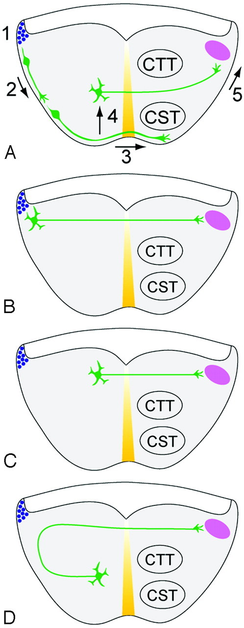
Conclusions: Our data suggest that the dorsal transverse axonal band in these disorders results from abnormal axonal pathfinding, abnormal neuronal migration, or a combination of the 2 processes.


Figures

Fig 1.
Fig 1.
A and B, Midsagittal T1-weighted images (T1WIs) in patients 1 and 3 show flattening of the ventral pons, thinning of the isthmus (arrowhead, A), dysmorphism of the dorsal upper pons (caplike) bulging (long arrows) and protruding in the fourth ventricle (B). The vermis is hypoplastic in A. C and D, Midsagittal T2WIs in patients 2 and 4 show the same findings as in A and B, with slightly different patterns (beaklike shape in D) (long arrow). In both cases, the vermis is hypoplastic and dysplastic and the CC is hypoplastic. The CC is dysmorphic in patient 3 (B) and thin in patient 4 (D). E and F, Midsagittal T1- and T2WIs for patients 5 and 6 show the tegmental cap with hypoplastic (E and F) and dysplastic (E) vermis. F, Fourth ventricle is slightly enlarged. The CC is hypoplastic in E and dysmorphic in F.
Fig 2.
Fig 2.
Patient 1: A and B, Coronal T1-weighted images and axial T2-weighted images (T2WIs) show the “molar tooth” appearance of the elongated SCPs running laterally (arrows) and the dorsal band crossing the midline and likely joining the MCPs (arrowheads). C, Axial T2WI at the level of the MCPs, which appear small (white arrows). D, Axial T2WI at the level of middle pons. A “horizontal cleft” is visible, outlined by black arrowheads. The black star indicates the fourth ventricle. Note the small size of cerebellar hemispheres (CH) and the hypoplastic vermis (V).
Fig 3.
Fig 3.
Patient 3: A and B, Color FA cross-sectional images show elongation of the SCP (green).The white star indicates the fourth ventricle. Descending long tracts (corticospinal and corticopontine) appear in blue. The ventral and middle transverse pontine fibers are missing; the SCP decussation is not visible. B, The ectopic bundle of fibers appears in red at the dorsal aspect of the pons and seems not to connect the MCPs visible laterally. C, Coronal FA image shows the elongated SCPs running laterally (arrows); the dorsal band (arrowheads) crossing the midline may join either the SCPs vertically or the MCPs horizontally. D and E, 3D projections of tractography. In the control (D), ventral transverse fibers are clearly seen (black arrow) and the MCPs are of normal size (arrowheads). In patient 3 (E), long descending tracts and the MCPs appear smaller (arrowheads). The ectopic dorsal pontine fibers are visible (long arrow) and are not seen to definitely connect the MCP (arrowheads).
Fig 4.
Fig 4.
Hypotheses to explain the aberrant dorsal position of transverse pontine fibers. A, Normal development. 1) Pontine gray neurons are produced from progenitors in the rhombic lip (blue circles). 2) Newly generated neurons migrate toward the ventral midline (yellow), the “anterior extramural migratory stream” of Altman and Bayer. 3) Axonal growth cones cross the ventral midline, leaving most neuronal cell bodies uncrossed. 4) Cell bodies migrate toward the ventricle along radial glia. 5) Axonal growth cones migrate into the MCP (pink). The transverse pontine fibers are sandwiched between the concurrently growing corticospinal tract (CST) and central tegmental tract (CTT). B, Hypothesis 1: Decreased ventral migration (step 2) leaves the pontine gray neurons scattered along the lateral pontine surface. Axonal growth cones cross the midline directly toward the contralateral MCP. C, Hypothesis 2: Increased radial migration (step 4) deposits pontine neurons near the ventricular surface of the pontine tegmentum. D, Hypothesis 3: Normal migration of pontine neurons. Note abnormal axon guidance away from the ventral surface, followed by midline crossing and attraction toward the MCP.

References

    1. Barkovich AJ, Millen KJ, Dobyns WB. A developmental classification of malformations of the brainstem. Ann Neurol 2007;62:625–39 - PubMed
    1. Maeoka Y, Yamamoto T, Ohtani K, et al. Pontine hypoplasia in a child with sensorineural deafness. Brain Dev 1997;19:436–39 - PubMed
    1. Ouanounou S, Saigal G, Birchansky S. Möbius syndrome. AJNR Am J Neuroradiol 2005;26:430–32 - PMC - PubMed
    1. Barth PG, Majoie CB, Caan MW, et al. Pontine tegmental cap dysplasia: a novel brain malformation with a defect in axonal guidance. Brain 2007;130 (pt 9):2258–66 - PubMed
    1. Tuch DS, Wisco JJ, Khachaturian MH, et al. Q-ball imaging Magn Reson Med 2004;52:1358–72 - PubMed

Publication types

LinkOut - more resources