. 2008 Nov;135(22):3643-53.
doi: 10.1242/dev.023325. Epub 2008 Oct 8.

Pioneer longitudinal axons navigate using floor plate and Slit/Robo signals

Affiliations

Pioneer longitudinal axons navigate using floor plate and Slit/Robo signals

W Todd Farmer et al. Development. 2008 Nov.

Abstract

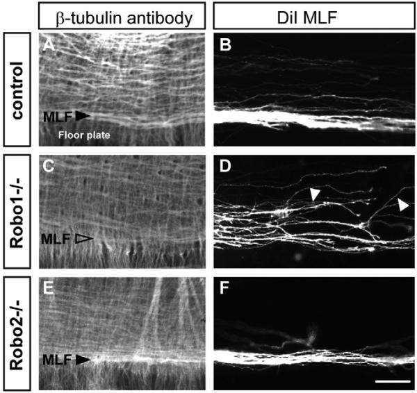
Longitudinal axons transmit all signals between the brain and spinal cord. Their axon tracts through the brain stem are established by a simple set of pioneer axons with precise trajectories parallel to the floor plate. To identify longitudinal guidance mechanisms in vivo, the overall role of floor plate tissue and the specific roles of Slit/Robo signals were tested. Ectopic induction or genetic deletion of the floor plate diverted longitudinal axons into abnormal trajectories. The expression patterns of the diffusible cues of the Slit family were altered in the floor plate experiments, suggesting their involvement in longitudinal guidance. Genetic tests of Slit1 and Slit2, and the Slit receptors Robo1 and Robo2 were carried out in mutant mice. Slit1;Slit2 double mutants had severe longitudinal errors, particularly for ventral axons, including midline crossing and wandering longitudinal trajectories. Robo1 and Robo2 were largely genetically redundant, and neither appeared to specify specific tract positions. However, combined Robo1 and Robo2 mutations strongly disrupted each pioneer tract. Thus, pioneer axons depend on long-range floor plate cues, with Slit/Robo signaling required for precise longitudinal trajectories.


Figures

Fig. 1
Fig. 1. Schematic of pioneer longitudinal tracts
The first neurons form an array of longitudinal axons with precise trajectories at specific DV positions (Easter et al., 1993; Mastick and Easter, 1996). The tracts include: ventral axons of the medial longitudinal fasciculus (MLF); dorsal axons of the lateral longitudinal fasciculus (LLF, originating from dorsal midbrain neurons, also known as the tract of the mesencephalic nucleus of the trigeminal nerve, tmesV); and a broad set of intermediate axons (termed here the ILF).
Fig. 2
Fig. 2. Sonic hedgehog transfection can divert longitudinal axon trajectories
A Sonic hedgehog (Shh) expression plasmid was co-electroporated with a GFP reporter plasmid into the hindbrain of 10- to 12-somite chick embryos. All panels show side views of whole hindbrain labels, anterior to left, dorsal to top. Axons were labeled with (A-C) DiI (red), with crystal locations shown in the insert of A, or (D) anti-β-tubulin (red). (A) Hindbrain of a Shh transfected embryo, as an open book with MLF and ILF axons labeled on both electroporated (top) and control sides. (B) Diffuse Shh electroporation showing both ventral and dorsal turning LLF responses. (C) Example of LLF axons turning towards the anterior (asterisks) near a Shh+ region. (D) Axonal antibody (red), showing reorientation of both longitudinal and commissural axon trajectories. (E) MLF axons (arrowheads) fan out within Shh-transfected patches. (F-I) Shh electroporation induces the ectopic floor plate markers Hnf3β and Slit1 (arrowheads); control (F,H) and electroporated hindbrains (G,I). fp, floor plate. Scale bars: 200 μm.
Fig. 3
Fig. 3. Loss of floor plate disrupts ipsilateral trajectories, dorsoventral position, and anteroposterior direction
(A,B,B′) Hindbrain of E10.5 control and Gli2–/– mouse embryos; open book whole mounts labeled with βIII tubulin antibody. Anterior to the left. In mutants, many axons project at angles and encroach into the midline. No MLF is visible. Dashed box enlarged in B′ highlights the mutant midline. (C,D) Axon tracing using DiI crystals placed at an intermediate position in r1 (letters in A and B). The midline is indicated by dashed lines. Control axons project posteriorly at intermediate positions, with a small number of commissural axons (CA). In mutants, many axons cross the midline and descend in bilaterally symmetrical tracts, with a few axons turning anteriorly. (Note that the DiI label site is similar in size and position in C and D, but appears larger in D because the mutant image in D was taken at a longer exposure, required to show the more widespread but less intensely labeled fibers.) (E,F) βIII tubulin antibody labeling of forebrain (FB) and midbrain (MB), side views of bisected whole mounts, truncated at the MB/HB boundary. In mutants, no MLF is visible, and several abnormal axon bundles are seen (arrowheads). (G-I) DiI tracing of projections from the ventral forebrain; side views of whole mounts. (H) MLF label of mutant shows no axons descending, but many axons ascending and crossing the midline. (I) Contralateral side of the same embryo, showing the anterior turning of abnormal post-crossing axons. (J-L) DiI tracing of dorsal midbrain axons; side views of whole midbrains. (K,L) Two mutant embryos, with numerous axons projecting at abnormal angles and/or anteriorly (arrowheads). The bar (J,K) indicates the distance between the descending LLF axons and the ventral midline. (C′,D′,G′-L′) Schematics of DiI labels. cf, cephalic flexure; FB, forebrain; HB, hindbrain; MB, midbrain; r2/r4, rhombomeres.
Fig. 4
Fig. 4. Changes in midline Slit expression correlate with axon guidance defects in Gli2 mutants
(A,B) βIII tubulin antibody labels in Gli2+/– and Gli2–/– embryos, showing the severe disruption of tracts. (C-H) In situ hybridization of three Slits, showing reduced Slit expression, with retention in r4 and the posterior hindbrain. In C, note the dorsal expression of low levels of Slit1 (see Fig. S1 in the supplementary material for midbrain expression of Slit1 and Slit2, but not Slit3). Scale bar: 200 μm.
Fig. 5
Fig. 5. Slit1 and Slit2 are required for many aspects of longitudinal axon guidance
Open-book hindbrain preparations of Slit1–/–;Slit2+/– and Slit1–/–;Slit2–/– E10.5 embryos, showing (A-C) βIII tubulin antibody and (D-O) DiI labeling of the LLF, ILF and MLF. Anterior, left; floor plate, down (or shown by dashed lines). (A,B) In Slit1–/–;Slit2–/– embryos, intermediate axons appear more diffuse, and the MLF is not visible. (C) Midline closeup showing longitudinal axons projecting in midbrain and anterior r1 floor plate. (D-F) DiI labels of LLF, from dorsal label sites in anterior r1. Most LLF axons in Slit1–/–;Slit2–/– mutants project normally, but a subset wander ventrally (arrowheads; E,F). (G-I) DiI labels of ILF, from intermediate anterior r1. In Slit1–/–;Slit2–/– mutants, the ILF appears shorter (H), and more axons wandering at divergent angles (I, arrowheads). (J-L) MLF labels from DiI placed adjacent to the floor plate in ventral midbrain. (K) Slit1–/–;Slit2+/– mutants show some axons that loop dorsally (arrowhead) in the anterior hindbrain. (L) The MLF in double mutants shows severe errors, including midline crossing and dorsal looping (arrowheads). (M-O) Slit1–/–;Slit2–/– double mutants. Midline sites back-label many MLF neurons, as well as bundles entering midbrain floor plate (N). (O) MLF axons project along the sides of hindbrain floor plate, crossing at multiple points. Some axons wander dorsally (arrowheads). These trajectory errors were verified by Robo1 antibody labeling (see Fig. S4 in the supplementary material). Scale bars: 200 μm in A,B,G,H,M; 50 μm in C,F,I,N; 100 μm in D,E,J-L,O.
Fig. 6
Fig. 6. Robo1 and Robo2 expression in longitudinal pioneer neurons
E10.5 whole mounts. (A,B) Differential expression of Robo1 and Robo2 mRNA by ISH. Insets show close-ups of midbrain with strong Robo1 signal in MLF cell bodies (arrowhead). Low signal is seen in the ILF for both Robo1 and Robo2 (dashed box). In the dorsal midbrain, tmesV cell bodies of LLF express Robo2 (dashed box). (C) Anti-Robo1 antibody labels axons of the MLF, and of the ILF, although at lower levels; anterior up. (D) X-gal labeling of homozygous Robo2 tau-lacZ knock-in embryos labels LLF axons (arrowheads) and possibly a subset of ILF, but not MLF.
Fig. 7
Fig. 7. Robo1 mutations alter MLF pathfinding
(A,B) Close-up views of the MLF in control embryos, showing a wild-type MLF bundle (A) and trajectories (B). (C,D) Robo1 mutants lack a distinct MLF bundle. (D) DiI labels show a widened MLF with some axons traversing a more dorsal position or taking a dorsal trajectory (arrowheads). (E,F) Robo2 mutants have a normal MLF (and other tracts). Scale bar: 50 μm.
Fig. 8
Fig. 8. Robo1 and Robo2 are required for longitudinal axon guidance
(A-C) Axonal antibody labels. Robo1–/–;Robo2–/– errors include axons diverging dorsally, a large number of axons in the floor plate, and disorganized intermediate axons. (C) Close up of dorsal looping axons in the posterior hindbrain. (D-F) Broad DiI labels of many longitudinal axons. Mutant trajectories are abnormal, tending to divert ventrally (arrowheads). (G-I) LLF labels, DiI in dorsal r1. (G) Control LLF projections parallel the bend in r2 tissue. (H,I) In Robo1–/–;Robo2–/– double mutant embryos, some LLF axons have a normal trajectory, but others make dorsal and ventral deviations (arrowheads). (J-L) ILF labels from r1. Few mutant ILF axons project straight longitudinally, and instead diverge dorsally or ventrally (arrowheads, K,L). (L) An ILF axon wandering dorsally then ventrally. Asterisks indicate the DiI label site. Scale bars: 200 μm in A,B; 50 μm in C,I,L; 100 μm in H,K (for G,J, respectively).
Fig. 9
Fig. 9. Robo1 and Robo2 are required for MLF guidance
(A-C) MLF labeling adjacent to the floor plate. A single DiI crystal was placed adjacent to the floor plate. (B) In Robo1–/–;Robo2–/– double mutants, DiI labels MLF axons that fan out and cross midline. (C) Close up of wandering axons. (D-F) DiI labeling in the midline of anterior r1 of Robo1–/–;Robo2–/– double mutants back-labels many MLF cell bodies and midline bundles. (The MLF cell bodies are faintly labeled in the example shown.) Note that numerous commissural axons (CA) were labeled in mutant embryos (D, arrowhead), but not in controls. (E) Closeup of back-labeled MLF cell bodies. Their axons project ventrally and bundle in the midline. (F) Closeup of axon bundles in the hindbrain floor plate, and diverging dorsal bundles. Scale bar: 200 μm in A,B,D; 50 μm in C,E,F.
Fig. 10
Fig. 10. Longitudinal axon trajectories in Gli2, Slit and Robo mutants
Schematic diagram of the axon trajectories, with thick and thin arrows indicating common and rare axon paths. Forks show where axons diverge from major bundles, not actual axon bifurcation. The floor plate is designated by the yellow box (residual in Gli2 mutants). In the Slit mutant diagram, the yellow box represents Slit3 expression.

Similar articles

Cited by

References

    1. Agarwala S, Ragsdale CW. A role for midbrain arcs in nucleogenesis. Development. 2002;129:5779–5788. - PubMed
    1. Agarwala S, Sanders TA, Ragsdale CW. Sonic hedgehog control of size and shape in midbrain pattern formation. Science. 2001;291:2147–2150. - PubMed
    1. Ahsan M, Riley KL, Schubert FR. Molecular mechanisms in the formation of the medial longitudinal fascicle. J. Anat. 2007;211:177–187. - PMC - PubMed
    1. Andrews W, Liapi A, Plachez C, Camurri L, Zhang J, Mori S, Murakami F, Parnavelas JG, Sundaresan V, Richards LJ. Robo1 regulates the development of major axon tracts and interneuron migration in the forebrain. Development. 2006;133:2243–2252. - PubMed
    1. Bagri A, Marin O, Plump AS, Mak J, Pleasure SJ, Rubenstein JL, Tessier-Lavigne M. Slit proteins prevent midline crossing and determine the dorsoventral position of major axonal pathways in the mammalian forebrain. Neuron. 2002;33:233–248. - PubMed

Publication types

MeSH terms