. 2008 Oct 8;28(41):10234-44.
doi: 10.1523/JNEUROSCI.3494-08.2008.

Ablation of proliferating microglia does not affect motor neuron degeneration in amyotrophic lateral sclerosis caused by mutant superoxide dismutase

Affiliations

Ablation of proliferating microglia does not affect motor neuron degeneration in amyotrophic lateral sclerosis caused by mutant superoxide dismutase

Genevíève Gowing et al. J Neurosci. .

Abstract

Microglial activation is a hallmark of all neurodegenerative diseases including amyotrophic lateral sclerosis (ALS). Here, a detailed characterization of the microglial cell population within the spinal cord of a mouse model of familial ALS was performed. Using flow cytometry, we detected three distinct microglial populations within the spinal cord of mice overexpressing mutant superoxide dismutase (SOD1): mature microglial cells (CD11b(+), CD45(low)), myeloid precursor cells (CD11b(+), CD45(int)), and macrophages (CD11b(+), CD45(high)). Characterization of cell proliferation within the CNS of SOD1(G93A) mice revealed that the expansion in microglial cell population is mainly attributable to the proliferation of myeloid precursor cells. To assess the contribution of proliferating microglia in motor neuron degeneration, we generated CD11b-TK(mut-30); SOD1(G93A) doubly transgenic mice that allow the elimination of proliferating microglia on administration of ganciclovir. Surprisingly, a 50% reduction in reactive microglia specifically in the lumbar spinal cord of CD11b-TK(mut-30); SOD1(G93A) doubly transgenic mice had no effect on motor neuron degeneration. This suggests that proliferating microglia-expressing mutant SOD1 are not central contributors of the neurodegenerative process in ALS caused by mutant SOD1.


Figures

Figure 1.
Figure 1.
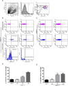
Expansion of the CD11b+ cell population, which consists of three different subpopulations, occurs early in the disease. A, Spinal cord (S.C.) sections of SOD1G93A and SOD1WT were stained for the myeloid marker CD11b. Gray matter immunopositive cells were counted at the age of 60, 80, 100, and 120 d for SOD1G93A and 130 d SOD1WT. *p < 0.05, ***p < 0.001, Significant differences versus SOD1WT control; p < 0.05, significant versus SOD1G93A at 60 d; †††p < 0.001, significant versus SOD1G93A 60 d; and ##p < 0.01, significant compared with SOD1G93A at 80 d. All values are mean ± SEM. B, Isolated spinal cord mononuclear cells were analyzed using 5-color flow cytometry. Cells were gated (R1, B1) using Annexin-5 and 7-AAD to eliminate apoptotic and necrotic cells (data not shown) and further analyzed for viability using 7-AAD (B2). Viable cells were analyzed for CD11b and CD45 expression (B3). Three CD11b+ populations were found: macrophages (orange, R4), myeloid precursor cells (purple, R3), and mature microglia (blue, R2). Because there are only few macrophages present in the mononuclear cell isolate, only myeloid precursor cells and mature microglia were further analyzed. C, Expression of the markers Gr1 and F4/80 in the different cellular subsets. Mature microglia were negative for both Gr1 and F4/80 (C1). Myeloid precursor cells were positive for Gr1 (C3) and negative for F4/80 (C3). Isotype stains are shown in C2 and C4, respectively. D, Contribution of macrophages (black), myeloid precursors (dark gray), and mature microglia (light gray) to the expansion of the CD11b+ cell population at different ages. At the age of 60 d, mature microglia are twice as numerous as the myeloid precursor cells (p < 0.05). At the age of 120 d, the proportion of myeloid precursor cells had greatly increased compared with the age of 130 d SODWT (p < 0.01). At 120 d, there were almost twice as many myeloid precursor cells than mature microglial cells present in the CD11b cell population (p < 0.0001). All values are mean ± SEM; n = 3–9 for all experiments.
Figure 2.
Figure 2.
Mature microglia obtain a dendritic cell phenotype as disease progresses. Five-color flow cytometric analysis of microglial cells using different markers of microglial activation. A, Isolated mononuclear spinal cord cells were gated (R1; A1) and viable cells (R1) selected using 7-AAD (P1; A2). A3, Cells were analyzed for CD11b/CD45 expression. Because there were only few macrophages present (R4, orange), we only analyzed myeloid precursor cells (purple, R3) and mature microglia (blue, R2). B, Myeloid precursor cells from SOD1G93A end-stage mice were CD11c negative (B1), whereas mature microglia (B3) were clearly positive for CD11c. B2, B4, Isotype controls for B1 and B3, respectively. B5, Fifty-nine percent of gated CD11c+ mature microglial cells coexpressed CD86. B6, Isotype control for B5. C, However, in SOD1WT mouse, both myeloid precursor cells (C1, compare with C2 isotype control) and mature microglia (C3, compare with C4 isotype control) did not express CD11c. D, E, Quantification of CD11c (D) and CD86 (E) expression of microglial cells at different ages both in SOD1G93A and SOD1WT mice. D, ***p < 0.0001 compared with SOD1WT at 130 d and SOD1G93A at 60 d and p < 0001 compared with SOD1G93A 80 d. E, **p < 0.01 compared with SOD1WT 130 d of age and SOD1G93A 60 d of age; all values are mean ± SEM. For CD11c expression, n = 3–13; for CD86 expression, n = 5–7.
Figure 3.
Figure 3.
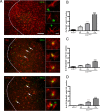
Proliferation in the SOD1G93A lumbar gray matter spinal cord. A, Double immunofluorescence for GFAP, NG2, or CD11b and BrdU in the lumbar spinal cord of SOD1G93A transgenic mice (arrows indicate double-labeled cells; dotted lines indicate the border between white matter and gray matter). Scale bars: 100 μm (insets); 10 μm. B–D, Quantification of cell proliferation at different ages in the SOD1G93A spinal cord compared with SOD1WT spinal cord. B, BrdU+ cells were counted at the ages of 80, 100, and 120 d in the SOD1G93A mouse and at 130 d in the SOD1WT mouse. ***p < 0.0001 compared with SOD1WT 130 d and SOD1G93A at 80 and 100 d; †††p < 0.0001 with SOD1WT 130 d; *p < 0.05 with SOD1G93A at 100 d and SOD1WT at 130 d. C–D, Number of BrdU cells double+ for NG2 (C) and CD11b (D) at the different ages examined. C, ***p < 0.0001 with SOD1WT 130 d and SOD1G93A at 80 d, and p < 0.001 with SOD1G93A 100 d. *p < 0.05 with SOD1WT. D, ***p < 0.0001 with SOD1WT 130 d and SOD1G93A at 80 d, and p < 0001 with SOD1G93A at 100 d. †††p < 0.001 with SOD1WT 130 d and p < 0.05 with SOD1G93A 80 d. All values are mean ± SEM; n = 4–9 for all experiments.
Figure 4.
Figure 4.
Ablation of proliferating microglia in TKmut-30; SOD1G93A transgenic mice. A, Schematic representation of mice receiving ganciclovir or saline intrathecally via an osmotic pump at L4–L5 spinal level. B, Immunohistochemistry for Iba1 and Mac-2 at the L5 level in TKmut-30; SOD1G93A or WT; SOD1G93A transgenic animals receiving ganciclovir. Magnification 20×. Scale bar, 100 μm. C–D, Quantifications of microglial cell ablation stained with Iba1 (C) or Mac-2 (D). *p < 0.05; ***p < 0.001. All values are mean ± SEM; n = 8–12 mice per analysis.
Figure 5.
Figure 5.
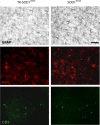
Influence of decreased microgliosis on astrocytes, glial progenitors, and T cell numbers. A, Immunohistochemistry for GFAP, NG2, and CD3 in the lumbar spinal cord of TKmut-30; SOD1G93A and WT; SOD1G93A. Magnification, 40×. Scale bar, 50 μm.

Similar articles

Cited by

References

    1. Ajami B, Bennett JL, Krieger C, Tetzlaff W, Rossi FM. Local self-renewal can sustain CNS microglia maintenance and function throughout adult life. Nat Neurosci. 2007;10:1538–1543. - PubMed
    1. Alexianu ME, Kozovska M, Appel SH. Immune reactivity in a mouse model of familial ALS correlates with disease progression. Neurology. 2001;57:1282–1289. - PubMed
    1. Almand B, Clark JI, Nikitina E, van Beynen J, English NR, Knight SC, Carbone DP, Gabrilovich DI. Increased production of immature myeloid cells in cancer patients: a mechanism of immunosuppression in cancer. J Immunol. 2001;166:678–689. - PubMed
    1. Austyn JM, Gordon S. F4/80, a monoclonal antibody directed specifically against the mouse macrophage. Eur J Immunol. 1981;11:805–815. - PubMed
    1. Banchereau J, Steinman RM. Dendritic cells and the control of immunity. Nature. 1998;392:245–252. - PubMed

Publication types

MeSH terms