Comparative Study
. 2008 Oct 8;28(41):10278-86.
doi: 10.1523/JNEUROSCI.2451-08.2008.

Distinctive features of adult ocular dominance plasticity

Affiliations
Comparative Study

Distinctive features of adult ocular dominance plasticity

Masaaki Sato et al. J Neurosci. .

Abstract

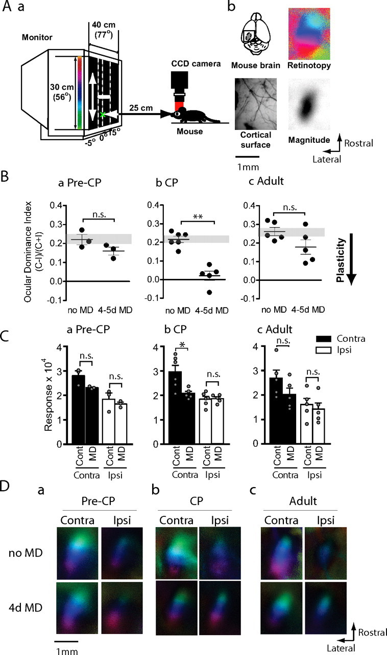
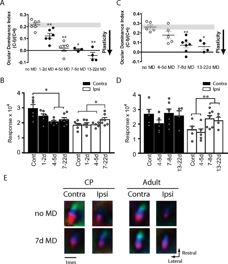
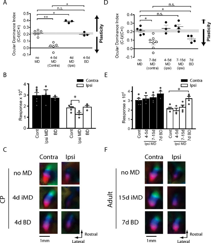
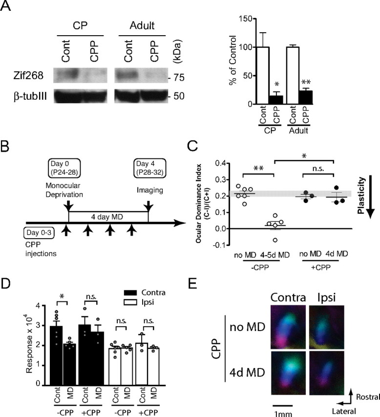
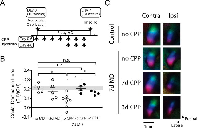
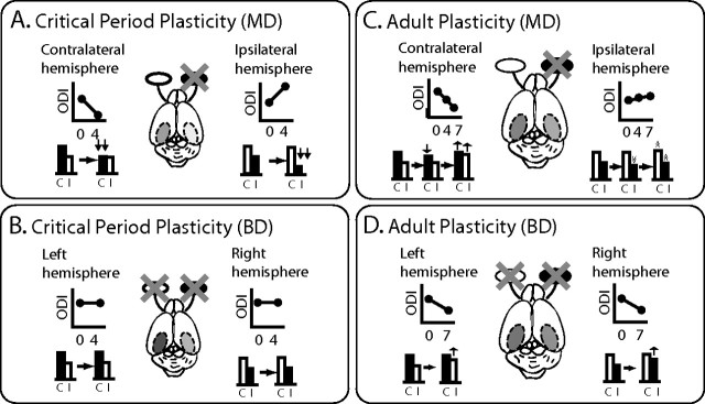
Sensory experience profoundly shapes neural circuitry of juvenile brain. Although the visual cortex of adult rodents retains a capacity for plasticity in response to monocular visual deprivation, the nature of this plasticity and the neural circuit changes that accompany it remain enigmatic. Here, we investigate differences between adult and juvenile ocular dominance plasticity using Fourier optical imaging of intrinsic signals in mouse visual cortex. This comparison reveals that adult plasticity takes longer than in the juvenile mouse, is of smaller magnitude, has a greater contribution from the increase in response to the open eye, and has less effect on the hemisphere ipsilateral to the deprived eye. Binocular deprivation also causes different changes in the adult. Adult plasticity is similar to juvenile plasticity in its dependence on signaling through NMDA receptors. We propose that adult ocular dominance plasticity arises from compensatory mechanisms that counterbalance the loss of afferent activity caused by visual deprivation.


Figures

Figure 1.
Figure 1.
The effects of brief MD on mice of different ages. Aa, A short sweeping bar moving upward or downward was presented in the binocular visual field of a mouse and the evoked cortical activity was imaged with a CCD camera. Ab, Occipital cortex of left hemisphere was exposed (top left) and cortical blood vessel pattern was captured under green light (bottom left). Activity maps of retinotopy (top right) and magnitude (bottom right) were imaged under red light to create polar maps as seen hereafter. Ba–Bc, OD index after brief (4–5 d) MD of the contralateral eye in precritical period mice (Ba, Pre-CP; n = 3 each), critical period mice (Bb, CP; n = 5–6) or adult mice (Bc, Adult; n = 5 each). The y-axis shows OD index as described in Materials and Methods. The mice subjected to brief MD (MD) showed a significantly lower OD index in CP, but neither in pre-CP nor adulthood. The downward arrow shown to right illustrates that a lower OD index after MD indicates greater plasticity. **p < 0.01. n.s., Not significant. Ca–Cc, Maximum response magnitude expressed as fractional change in light reflectance elicited by stimulation of the contralateral (black bars) or ipsilateral (white bars) eye of mice deprived before (Ca) or during (Cb) the critical period or in adulthood (Cc). Brief MD reduced responsiveness of the deprived eye significantly only in CP, but not pre-CP or in adulthood. *p < 0.05. Da–Dc, Polar maps of responses in binocular visual cortex of mice of different ages with or without brief MD.
Figure 2.
Figure 2.
Critical period ocular dominance plasticity and adult ocular dominance plasticity show different time courses and response changes. A, Time course of OD shifts in the critical period after varying lengths of MD. Data indicated by open circles were taken from Figure 1 for comparison (n = 3–6; *p < 0.05, **p < 0.01). B, Maximum magnitude of responses elicited by the stimulus to the contralateral (Contra; black bars) or ipsilateral (Ipsi; white bars) after varying length of MD in critical period mice. For statistical comparison, data from groups that were qualitatively indistinguishable were pooled as indicated (n = 6–17; *p < 0.05). Cont, Control. C, Time course of OD shift in adult mice after varying length of MD. Data indicated in open circles were taken from Figure 1 for comparison (n = 4–7; *p < 0.05, **p < 0.01). D, Maximum magnitude of responses elicited by the stimulus to the contralateral (black bars) or ipsilateral eye (white bars) in adult mice. Data from groups that did not show qualitative difference were pooled as indicated for statistical comparison (n = 10–11; **p < 0.01). E, Polar maps of cortical responses in critical period and adult mice with or without 7 d MD.
Figure 3.
Figure 3.
Effects of ipsilateral MD and BD in juvenile and adult mice. A, OD shifts after 4 d ipsilateral-eye MD and BD in critical period mice. The OD bias shifted toward the contralateral nondeprived eye after 4 d ipsilateral MD. BD did not change OD (n = 3–6; *p < 0.05, **p < 0.01). Data indicated in open circles were taken from Figure 1 for comparison. B, Maximum response magnitude for each eye after 4 d ipsilateral-eye MD and BD. The value for the ipsilateral closed eye response of mice with 4 d ipsilateral MD was significantly smaller than that of control mice (*p < 0.05, Cont Ipsi vs Ipsi MD Ipsi; n = 3–6). C, Polar maps of cortical responses in critical period mice with no MD, 4 d ipsilateral MD, and BD. D, OD after 7–15 d ipsilateral-eye MD and 7 d BD in adult mice. OD changed only very slightly toward the contralateral nondeprived eye after ipsilateral-eye MD. BD caused moderate and significant ODI shift toward the ipsilateral eye (n = 3–7; *p < 0.05). Data indicated in open circles were taken from Figure 1 for comparison. E, Maximum response magnitude for each eye after 7–15 d ipsilateral MD and 7 d BD in adult mice. The response magnitude of the ipsilateral eye after BD is significantly larger than that of mice without deprivation (n = 4–5; *p < 0.05). F, Polar maps of cortical responses in adult mice with no MD, 7 d ipsilateral MD, and BD. Contra, Contralateral; Ipsi, ipsilateral.
Figure 4.
Figure 4.
CPP-mediated suppression of NMDA receptor function in visual cortex and NMDA receptor-dependent ocular dominance plasticity in the critical period. A, Suppression of Zif268 expression in visual cortex at 24 h after CPP injection in juvenile and adult mice. CPP or control saline (Cont) were injected into mice in the critical period at P28 and in adulthood at P80 and the amount of zif268 (top) and β III tubulin (bottom) proteins at 24 h after injection was quantified by immunoblotting. The right panel shows summary data (n = 3 each; *p < 0.05, **p < 0.01). B, Mice in the critical period were treated with CPP 4–6 h before MD on day 0 and every 24 h thereafter and were imaged at 4 d after MD. C, OD shifts after 4 d MD were blocked by daily CPP treatment in critical period mice (n = 3–6; *p < 0.05, **p < 0.01). Data indicated in open circles were taken from Figure 1 for comparison. D, Decrease in the response of the deprived eye after 4 d MD was prevented by daily CPP treatment in critical period mice (n = 3–6). E, Polar maps of cortical responses of critical period mice with or without CPP treatment and MD. Contra, Contralateral; Ipsi, ipsilateral.
Figure 5.
Figure 5.
Role of NMDA receptor activation in adult ocular dominance plasticity. A, In 7 d CPP treatment (days 0–6), adult mice were treated with CPP 4–6 h before MD on day 0 and every 24 h thereafter, and were imaged at 7 d after MD. In 3 d CPP treatment (days 4–6), adult mice were subjected to 7 d MD with no CPP treatment for the first 4 d. Daily CPP injection was then started from day 4 to day 6. B, OD shifts after 7 d MD in adult mice were fully blocked by 7 d CPP treatment (n = 4–7; *p < 0.05). Mice with 3 d CPP treatment showed partial blockade of OD shifts compared with the full effect produced by 7 d MD (n = 4–7; *p < 0.05). Data indicated in open circles were taken from Figures 1 and 2 for comparison. C, Polar maps of cortical responses in adult mice with or without CPP treatment and MD. Contra, Contralateral; Ipsi, ipsilateral.
Figure 6.
Figure 6.
Different functional consequences for binocular responsiveness in the critical period and adult visual cortices after monocular and binocular deprivation. A, In juvenile mice, brief (4 d) MD causes robust shift in ocular dominance toward the open eye in both the contralateral and ipsilateral hemispheres. These processes are mediated primarily by rapid decreases in the responsiveness of the deprived eyes. The degree of plasticity is equal in the two hemispheres. B, BD has no effect on ocular dominance, suggesting that OD shifts produced by brief MD in the critical period are based on competition between inputs from the two eyes. C, In contrast, in adult mice, longer (7 d) MD causes a significantly large shift in the contralateral hemisphere to the deprived eye and only a hint of OD shift in the ipsilateral hemisphere. These shifts are mediated by transient small decreases in the response of the deprived eye at 4 d after MD followed by recovery from the depression of the response of the deprived eye and an increase in the response to the nondeprived eye at 7 d after MD. This asymmetry suggests that the degree of plasticity is determined by the strength of the deprived input. D, BD (7 d) causes a mild and atypical OD shift. This implies that the OD shifts in adult visual cortex are not based purely on competition in the conventional sense.

References

    1. Antonini A, Fagiolini M, Stryker MP. Anatomical correlates of functional plasticity in mouse visual cortex. J Neurosci. 1999;19:4388–4406. - PMC - PubMed
    1. Buonomano DV, Merzenich MM. Cortical plasticity: from synapses to maps. Annu Rev Neurosci. 1998;21:149–186. - PubMed
    1. Cang J, Kalatsky VA, Löwel S, Stryker MP. Optical imaging of the intrinsic signal as a measure of cortical plasticity in the mouse. Vis Neurosci. 2005;22:685–691. - PMC - PubMed
    1. Daw NW, Fox K, Sato H, Czepita D. Critical period for monocular deprivation in the cat visual cortex. J Neurophysiol. 1992;67:197–202. - PubMed
    1. Fischer QS, Graves A, Evans S, Lickey ME, Pham TA. Monocular deprivation in adult mice alters visual acuity and single-unit activity. Learn Mem. 2007;14:277–286. - PMC - PubMed

Publication types

Substances

LinkOut - more resources