. 2008 Oct 8;28(41):10339-48.
doi: 10.1523/JNEUROSCI.1917-08.2008.

The Reelin signaling pathway promotes dendritic spine development in hippocampal neurons

Affiliations

The Reelin signaling pathway promotes dendritic spine development in hippocampal neurons

Sanyong Niu et al. J Neurosci. .

Abstract

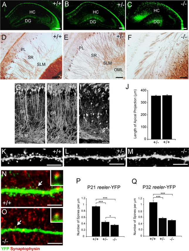
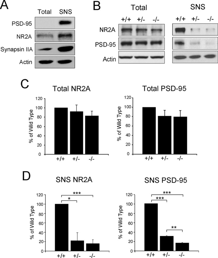
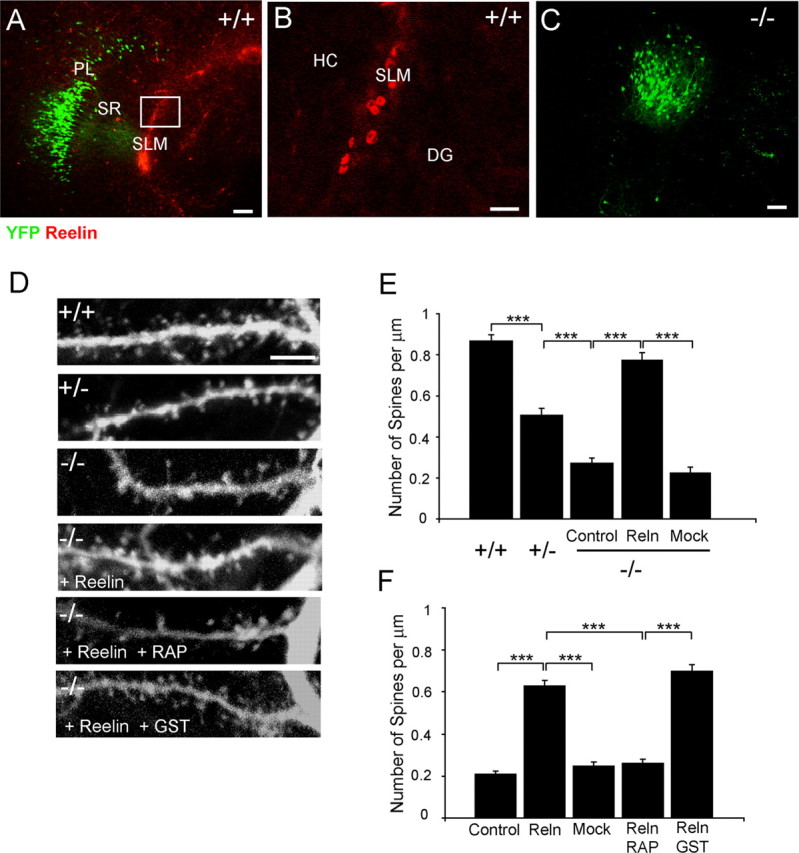
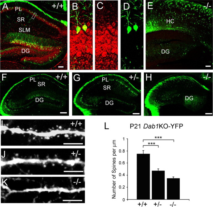
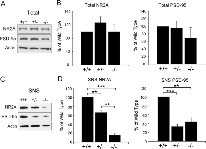
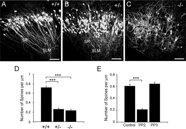
The development of distinct cellular layers and precise synaptic circuits is essential for the formation of well functioning cortical structures in the mammalian brain. The extracellular protein Reelin, through the activation of a core signaling pathway, including the receptors ApoER2 and VLDLR (very low density lipoprotein receptor) and the adapter protein Dab1 (Disabled-1), controls the positioning of radially migrating principal neurons, promotes the extension of dendritic processes in immature forebrain neurons, and affects synaptic transmission. Here we report for the first time that the Reelin signaling pathway promotes the development of postsynaptic structures such as dendritic spines in hippocampal pyramidal neurons. Our data underscore the importance of Reelin as a factor that promotes the maturation of target neuronal populations and the development of excitatory circuits in the postnatal hippocampus. These findings may have implications for understanding the origin of cognitive disorders associated with Reelin deficiency.


Figures

Figure 1.
Figure 1.
Dendritic spine density is reduced in hippocampal pyramidal neurons of heterozygous and homozygous reeler mice. A–C, Low-magnification confocal images of the hippocampus of adult wild-type, heterozygous, or homozygous reeler-YFP littermates. YFP-positive neurons are ectopic in homozygous reeler mice. Scale bars, 200 μm. D–F, YFP immunohistochemistry in floating sections of wild-type, heterozygous, or homozygous reeler-YFP littermates. Scale bars, 100 μm. G–I, Composite confocal images of CA1 apical projections of adult wild-type, heterozygous, or homozygous reeler-YFP littermates. YFP-positive pyramidal neurons appear normal in heterozygous mice, whereas they are stunted and misoriented in homozygous reeler mice. Scale bars, 50 μm. J, Quantification of the extent of the apical projection in wild-type and heterozygous reeler mice reveal no difference between these genotypes. n = 15 measurements per genotype. Bar graphs show the mean ± SEM. K–M, High-magnification representative confocal images of second-order apical dendrite branches of CA1 pyramidal neurons. Mature spines are present in all genotypes. Scale bars, 5 μm. N–O, Confocal images of wild-type or reeler mutant YFP-positive dendrites double labeled with synaptophysin antibodies. The insets show enlarged images of the synapses indicated by the arrows. Scale bars, 5 μm. P–Q, Quantification of spine density in second- to fourth-order apical dendrite branches in P21 (P) or P32 (Q) mice. Spine density is significantly reduced in heterozygous and homozygous reeler compared with wild-type mice at both ages. n = 25 dendrite segments per genotype from three independent experiments. Bar graphs show the mean ± SEM; *p < 0.03; ***p < 0.001. HC, hippocampus proper; DG, dentate gyrus; PL, pyramidal layer.
Figure 2.
Figure 2.
Reduced expression and synaptic localization of postsynaptic proteins in the hippocampus of heterozygous and homozygous reeler mice. A, Western blot analysis of presynaptic and postsynaptic proteins in total extracts and crude synaptosomal (SNS) fractions of wild-type mouse hippocampus. Postsynaptic proteins PSD-95 and NR2A, and the presynaptic protein synapsin IIA are enriched in SNS fractions. Blots were reprobed with actin antibodies as a protein-loading control. B, Western blot analysis of postsynaptic proteins in total extracts and SNS fractions of wild-type, heterozygous, and homozygous reeler hippocampi. Quantification of the data is from triplicate experiments. C, D, Total levels of NR2A and PSD-95 are similar (C), whereas SNS levels (D) are significantly reduced in heterozygous and homozygous reeler mice compared with wild type.
Figure 3.
Figure 3.
Reelin rescues the dendritic spine density deficit in reeler hippocampal slice cultures in a manner that is dependent on the activity of lipoprotein receptors. A–C, Double labeling of organotypic slice cultures obtained from YFP-positive wild-type or homozygous reeler hippocampi. Direct YFP fluorescence (green) and Reelin immunofluorescence (red) are shown. B is an enlargement of the boxed area in A. Reelin is expressed by Cajal-Retzius cells in the SLM. PL, pyramidal layer; HC, hippocampus proper; DG, dentate gyrus. Scale bars: A, C, 100 μm; B, 20 μm. D, Representative confocal images of second-order apical dendrite branches of CA1 pyramidal neurons in hippocampal cultures obtained from wild-type, heterozygous, or homozygous reeler mice and treated as indicated. More mature spines are present in wild-type cultures and in mutant cultures treated with Reelin and Reelin plus control GST, but not Reelin plus RAP. Scale bars, 5 μm. E, Quantification of spine density in second- to fourth-order apical dendrite branches in wild-type, heterozygous, or homozygous reeler hippocampal slice cultures. Spine density is significantly reduced in heterozygous and homozygous reeler compared with wild-type cultures. Spine density is significantly increased in mutant cultures treated with Reelin compared with mock-treated or untreated (control) slices. F, Quantification of spine density in second- to fourth-order apical dendrite branches from homozygous reeler hippocampal slices left untreated (control), treated with Reelin-conditioned medium, mock medium, Reelin plus RAP, or Reelin plus GST control. n = 20–35 dendrite segments per genotype or treatment from three independent experiments. Bar graphs (E, F) show the mean ± SEM; ***p < 0.001.
Figure 4.
Figure 4.
Dendritic spine density is reduced in hippocampal pyramidal neurons of heterozygous and homozygous Dab1KO-YFP mice. A–E, Double labeling of YFP (green) and Dab1 (red) immunoreactivity in hippocampal sections from wild-type (A–D) and homozygous Dab1 knock-out mice (E). Images in B–D are enlargements of the boxed area in A. Dab1 is expressed in YFP-positive pyramidal neurons in wild-type hippocampus. PL, pyramidal layer; HC, hippocampus proper; DG, dentate gyrus. Scale bars: A, E, 100 μm. F–H, Low-magnification confocal images of the hippocampus of adult YFP-positive wild-type, heterozygous, or homozygous Dab1KO-YFP littermates. YFP-positive neurons are ectopic in homozygous Dab1 knock-out mice. Scale bars, 200 μm. I–K, Representative images of second-order apical dendrite branches of YFP-positive CA1 pyramidal neurons. Mature spines are present in all genotypes. Scale bars, 5 μm. L, Quantification of spine density in second- to fourth-order apical dendrite branches. Spine density is significantly reduced in heterozygous and homozygous Dab1 knock-out compared with wild-type mice. n = 25 dendrite segments per genotype from three independent experiments. Bar graphs show the mean ± SEM; ***p < 0.001.
Figure 5.
Figure 5.
A–D, Reduced synaptic localization of postsynaptic proteins in the hippocampus of heterozygous and homozygous Dab1KO mice. Western blot analysis of postsynaptic proteins in total protein extracts (A) or synaptosomal (SNS) fractions (C) from the hippocampi of wild-type, heterozygous, and homozygous Dab1 knock-out mice was performed. Blots were probed with NR2A and PSD-95 antibodies and, subsequently, with actin antibodies as a protein loading control. Quantification of the data was from three Western blot experiments. Total levels (B) of NR2A and PSD-95 were not significantly different. SNS levels (D) were significantly reduced in heterozygous and homozygous Dab1 knock-out mice compared with wild-type littermates. Bar graphs show the mean ± SEM. **p < 0.01, ***p < 0.001.
Figure 6.
Figure 6.
Dab1 and SFK activity are required for the development of a normal dendritic spine density in organotypic hippocampal cultures. A–C, Organotypic cultures of the hippocampus of wild-type, heterozygous, and homozygous Dab1KO-YFP littermates. YFP-positive neurons are disorganized in the mutant cultures. PL, pyramidal layer. Scale bars, 100 μm. D–E, Quantification of spine density in second- to fourth-order apical dendrite branches in Dab1KO wild-type, heterozygous, or homozygous littermates (D) or branches of wild-type slice cultures treated with the PP2 SFK inhibitor or the inert control PP3 (E). Spine density is significantly reduced in heterozygous and homozygous Dab1 knock-out compared with wild-type mice. n = 19–27 dendrite segments per genotype from three independent experiments. Spine density is also significantly decreased in PP2-treated cultures compared with controls. n = 24–30 dendrite segments per treatment from three independent experiments. Bar graphs show the mean ± SEM. ***p < 0.001.

Similar articles

Cited by

References

    1. Alcántara S, Ruiz M, D'Arcangelo G, Ezan F, de Lecea L, Curran T, Sotelo C, Soriano E. Regional and cellular patterns of reelin mRNA expression in the forebrain of the developing and adult mouse. J Neurosci. 1998;18:7779–7799. - PMC - PubMed
    1. Arnaud L, Ballif BA, Förster E, Cooper JA. Fyn tyrosine kinase is a critical regulator of disabled-1 during brain development. Curr Biol. 2003;13:9–17. - PubMed
    1. Ballif BA, Arnaud L, Cooper JA. Tyrosine phosphorylation of Disabled-1 is essential for Reelin-stimulated activation of Akt and Src family kinases. Brain Res Mol Brain Res. 2003;117:152–159. - PubMed
    1. Beffert U, Weeber EJ, Durudas A, Qiu S, Masiulis I, Sweatt JD, Li WP, Adelmann G, Frotscher M, Hammer RE, Herz J. Modulation of synaptic plasticity and memory by Reelin involves differential splicing of the lipoprotein receptor ApoER2. Neuron. 2005;47:567–579. - PubMed
    1. Bock HH, Herz J. Reelin activates SRC family tyrosine kinases in neurons. Curr Biol. 2003;13:18–26. - PubMed

Publication types

MeSH terms

LinkOut - more resources