Comparative Study
. 2008 Oct 8;28(41):10386-98.
doi: 10.1523/JNEUROSCI.2387-08.2008.

GABAergic afferents activate both GABAA and GABAB receptors in mouse substantia nigra dopaminergic neurons in vivo

Affiliations
Comparative Study

GABAergic afferents activate both GABAA and GABAB receptors in mouse substantia nigra dopaminergic neurons in vivo

Elena Brazhnik et al. J Neurosci. .

Abstract

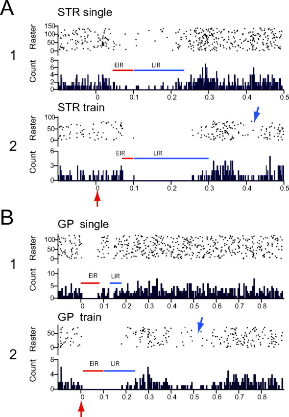
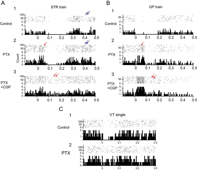
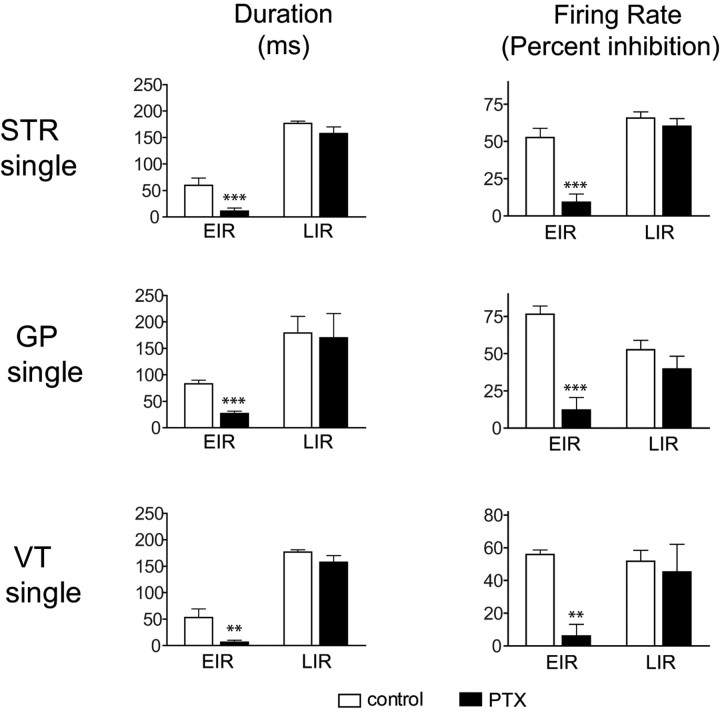
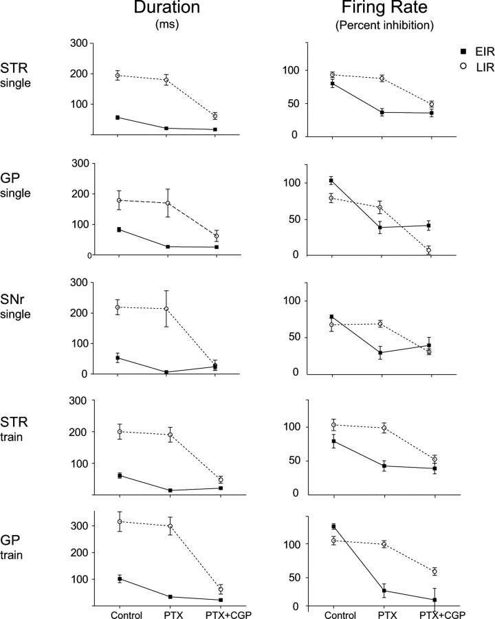
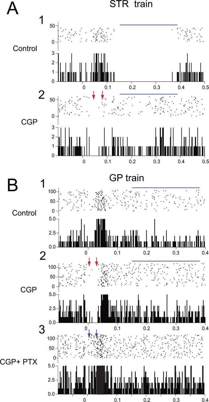
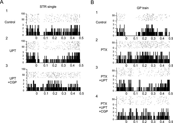
Most in vivo electrophysiological studies of substantia nigra have used rats. With the recent proliferation of the use of mice for in vitro neurophysiological studies because of the availability of various genetically modified strains to identify the roles of various channels and proteins in neuronal function, it is crucial to obtain data on in vivo responses in mice to verify that the in vitro results reflect functioning of systems comparable with those that have been well studied in rat. Inhibitory responses of rat nigral dopaminergic neurons by stimulation of afferents from striatum, globus pallidus, or pars reticulata have been shown to be mediated predominantly or exclusively by GABA(A) receptors. This is puzzling given the substantial expression of GABA(B) receptors and the ubiquitous appearance of GABA(B) synaptic responses in rat dopaminergic neurons in vitro. In the present study, we studied electrically evoked GABAergic inhibition in nigral dopaminergic neurons in C57BL/6J mice. Stimulation of the three major GABAergic inputs elicited stronger and longer-lasting inhibitory responses than those seen in rats. The early inhibition was GABA(A) mediated, whereas the later component, absent in rats, was GABA(B) mediated and selectively enhanced by GABA uptake inhibition. Striatal-evoked inhibition exhibited a slower onset and a weaker initial component compared with inhibition from globus pallidus or substantia nigra pars reticulata. These results are discussed with respect to differences in the size and neuronal density of the rat and mouse brain and the different sites of synaptic contact of the synapses from the three GABAergic afferents.


Figures

Figure 1.
Figure 1.
Representative extracellular single-unit recordings from dopaminergic neurons in vivo in mouse substantia nigra. A, Ten consecutive sweeps showing antidromic responses of a nigral neuron from striatal stimulation. Note that approximately half of the responses consist of the initial segment spike only (green traces), and that, although the initial segment latency is extremely constant, the somatodendritic component of the antidromic spike exhibits considerable latency variability (black traces). A collision occurs in the red trace. B, Ten consecutive spontaneous spikes (high-pass filtered) superimposed from a neuron firing in the pacemaker mode exhibit the long-duration waveform of nigrostriatal dopaminergic neurons commonly seen in rats. C, Five superimposed spontaneous spikes (high-pass filtered and 10 consecutive spikes averaged) from a neuron firing in the bursting mode exhibits the prominent initial segment–somatodendritic break (arrow) sometimes seen in spontaneous spikes as well as in antidromic responses. D–F, Samples of spontaneous activity from control neurons showing that mouse nigrostriatal neurons exhibit the same three patterns of firing seen in rat dopaminergic neurons: pacemaker (D), random (E), and bursty (F). Each top panel consists of 10 s of spontaneous activity with the corresponding autocorrelogram shown below. Autocorrelograms constructed from 750–100 spontaneous spikes. Bin width, 5 ms. G, Twenty-five consecutive sweeps superimposed to show a typical inhibitory response of a nigrostriatal neuron to single-pulse striatal stimulation (400 μA). Note the long latency to the onset of inhibition, the incomplete nature of the spike suppression, and the long-lasting inhibition. H, Twenty-five consecutive sweeps superimposed to show a typical inhibitory response of a nigrostriatal neuron to single-pulse stimulation of GP (500 μA). Note the rapid onset of the inhibition and the complete suppression of firing during the inhibitory period that is shorter than that evoked from striatum.
Figure 2.
Figure 2.
A, B, Effects of single-pulse stimulation and short trains of pulses (arrows) delivered to striatum (A; STR) and GP (B) on the spontaneous activity of one typical mouse nigral dopaminergic neuron in vivo. Note different timescales for striatal and pallidal responses. Inhibitory responses between time 0 and 100 ms were termed the early inhibitory responses (red lines), whereas those with onset >100 ms were termed the late inhibitory responses (blue lines). Note that the EIR evoked from striatal stimulation has a delayed onset and, in the case of single-pulse stimulation in A1, the firing is not completely suppressed. Concurrently, in both A1 and A2, there is a pronounced LIR that, in the case of train stimulation, comprises complete suppression of firing. In contrast, in response to GP stimulation, the EIR exhibits a very short-onset latency and comprises complete suppression of firing. In response to single-pulse stimulation in B1, the LIR is brief, but, after train stimulation, the LIR is of longer duration and also comprises complete suppression of firing. Blue arrows indicate examples of a secondary, very long-latency inhibition occasionally observed. In this and all subsequent PSTHs, the stimulus is delivered at time 0 (red arrow). Each PSTH consists of 75–100 trials. Bin width, 2 ms.
Figure 3.
Figure 3.
Summary graphs of the EIR and LIR of dopaminergic neurons to single-pulse and train stimulation of the striatum (STR), GP, and SNr. A, Both single-pulse and train stimulation of striatum typically evoked the EIR and the LIR, with a small proportion of cells responding with the EIR only. Train stimulation significantly increased the duration and the strength of the LIR compared with single stimuli. B, More than 70% of the neurons responded to single-pulse GP stimuli with EIR-only responses, whereas train stimulation elicited both the EIR and the LIR. Train stimulation in GP is extraordinarily effective at increasing the duration and strength of the evoked inhibition. C, Stimulation of SNr has a profile similar to that of GP in which a large proportion of neurons respond to single-pulse stimuli only with EIR. However, train stimulation evoked the LIR in 100% of the small sample of neurons tested. Train effects on duration and strength of inhibition were not significantly different from single pulse, probably because of the small n. *p < 0.05 and ***p < 0.001 compared with single pulse. STR, n = 37 neuron; GP, n = 44 neurons; and SNr, n = 3 neurons. VT, Ventral thalamus.
Figure 4.
Figure 4.
Typical PSTHs illustrating the effects of local blockade of GABAA and/or GABAB receptors on stimulation-induced inhibition. A1, Striatal train stimulation elicits an EIR and an LIR. Note delayed period of inhibition at ∼400 ms (double blue arrows). A2, Local application of PTX blocks the EIR, unmasking a brief excitation (red arrow; for explanation, see Results) and eliminates the delayed inhibition (double blue arrows) but does not affect the LIR. Note that the overall firing rate is increased. A3, Subsequent simultaneous application of PTX and CGP greatly attenuates the LIR (double red arrows), and the EIR remains blocked. Note that the overall firing rate is greater than in control but less than with PTX alone in A2. B1, A brief train to GP with same parameters as in A evokes near-complete suppression of firing during both the EIR and LIR. B2, Local application of PTX completely blocks the EIR, leaving the same excitation as seen in A2 (red arrow) but the LIR remains intact. B3, Subsequent combined application of PTX and CGP eliminates both the LIR (double red arrow) and the EIR. C1, SNr stimulation produces a short-latency EIR exhibiting complete suppression of activity. C2, Application of PTX completely abolishes the EIR. Note the absence of the excitatory response that is evoked by striatal or pallidal stimulation during GABAA receptor blockade. Stimulus is delivered at time 0. Each PSTH consists of 75–100 trials. Bin width, 2 ms. VT, Ventral thalamus.
Figure 5.
Figure 5.
Summary histograms displaying the effects of local application of PTX on the duration and firing rate inhibition of the EIR and LIR evoked by single-pulse stimulation of striatum, GP, and SNr. Note that the duration of the EIR and the magnitude of the inhibition of the EIR are significantly reduced, but there is no significant effect on the LIR, indicating that the EIR is predominantly or exclusively GABAA-mediated whereas the LIR is not. *p < 0.05 and ***p < 0.001 compared with baseline. VT, Ventral thalamus.
Figure 6.
Figure 6.
Summary histograms displaying the effects of local application of CGP on mean ± SEM duration and magnitude of the EIR and LIR evoked by train pulse stimulation of striatum and GP and single-pulse stimulation of SNr. Neither the duration or the magnitude of inhibition of the EIR is affected, but there is significant reduction of both duration and depression in firing rate from all three sites in the LIR, indicating that the LIR contains a significant GABAB-mediated component whereas the EIR does not. **p < 0.01 and ***p < 0.001 compared with baseline. VT, Ventral thalamus.
Figure 7.
Figure 7.
Summary of the effects of local application of PTX and CGP on the EIR and LIR. Means ± SEM duration and magnitude of inhibition (percentage inhibition, right) for both EIR (black circles, solid line) and LIR (white circles, dashed line) are shown for control condition (Control), after local administration of PTX, and after application of PTX together with CGP (PTX+CGP). STR, Striatum.
Figure 8.
Figure 8.
Presynaptic effect of GABAB receptor blockade increases EIR but blocks LIR in two representative neurons. A1, Control trace showing a modest EIR but a strong LIR (blue line) to striatal train stimulation. A2, Local application of CGP greatly increases the strength and duration of the EIR (red arrows) but at the same time almost completely abolishes the LIR (blue line). B1, Control trace in a second neuron showing relatively minimal EIR with incomplete suppression of activity and a more prominent LIR to GP train stimulation. B2, Local application of CGP greatly increases the strength and duration of the EIR, unmasking a period of complete suppression of spiking (red arrows) but also completely abolishing the LIR. B3, Subsequent application of both CGP and PTX abolishes the unmasked EIR (blue arrows).
Figure 9.
Figure 9.
Effects of GABAA and GABAB antagonists on spontaneous activity of mouse nigral dopaminergic neurons. A, Under control conditions, mouse dopaminergic neurons fire spontaneously at 4.6 spikes/s, approximately the same rate as in rats. Local application of PTX significantly increased firing rate to 6.2 spikes/s. Conversely, CGP produced a modest but statistically significant decrease in firing rate from 5.4 to 4.7 spikes/s, whereas combined application of CGP and PTX was similar to the effects of PTX alone and significantly increased spontaneous firing rate from 5.2 to 6.2 spikes/s. B, PTX also exerts dramatic effects on firing pattern, shifting the distribution to predominantly bursty firing and essentially eliminating the pacemaker mode. CGP exerts the opposite effect, decreasing burst firing and more than doubling the proportion of neurons firing in the pacemaker mode. Combined application of PTX and CGP produces effects qualitatively similar to those of PTX alone but with smaller proportional shifts in firing pattern. Numbers below the bars indicate the number of neurons. **p < 0.01; ***p < 0.001 (Bonferroni's t test); 111p < 0.001; 1p < 0.01 (χ2 test).
Figure 10.
Figure 10.
Blockade of GABA uptake with systemic administration of NO-711 (UPT) clearly enhances the LIR. A1, Baseline data show a clear EIR to single-pulse striatal stimulation with little or no LIR apparent. A2, After local application of UPT, the EIR is marginally enhanced, but now there is also a very clear LIR. A3, Combined application of UPT and CGP has a minimal attenuating effect on the EIR but completely blocks the increased LIR induced by uptake blockade. B1, Baseline trace showing complete suppression of firing during the EIR in response to train stimulation from GP. B2, Local application of PTX completely abolishes all inhibition. B3, Subsequent administration of NO-711 reveals a previously unseen, very pronounced LIR. B4, Subsequent application of CGP in the presence of PTX and GABA uptake blockade completely abolishes the LIR.

References

    1. Aosaki T, Graybiel AM, Kimura M. Effect of the nigrostriatal dopamine system on acquired neural responses in the striatum of behaving monkeys. Science. 1994;265:412–415. - PubMed
    1. Bishop KM, Wahlsten D. Sex and species differences in mouse and rat forebrain commissures depend on the method of adjusting for brain size. Brain Res. 1999;815:358–366. - PubMed
    1. Bolam JP, Smith Y. The GABA and substance P input to dopaminergic neurones in the substantia nigra of the rat. Brain Res. 1990;529:57–78. - PubMed
    1. Borden LA, Murali Dhar TG, Smith KE, Weinshank RL, Branchek TA, Gluchowski C. Tiagabine, SK&F 89976-A, CI-966, and NNC-711 are selective for the cloned GABA transporter GAT-1. Eur J Pharmacol. 1994;269:219–224. - PubMed
    1. Bowery NG, Hudson AL, Price GW. GABAA and GABAB receptor site distribution in the rat central nervous system. Neuroscience. 1987;20:365–383. - PubMed

Publication types

MeSH terms

LinkOut - more resources