. 2008 Dec;100(6):3264-74.
doi: 10.1152/jn.90418.2008. Epub 2008 Oct 8.

BDNF modulation of NMDA receptors is activity dependent

Affiliations

BDNF modulation of NMDA receptors is activity dependent

Robert A Crozier et al. J Neurophysiol. 2008 Dec.

Abstract

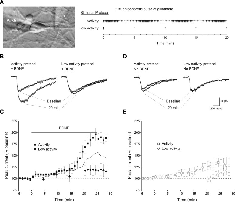
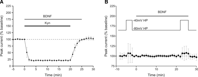
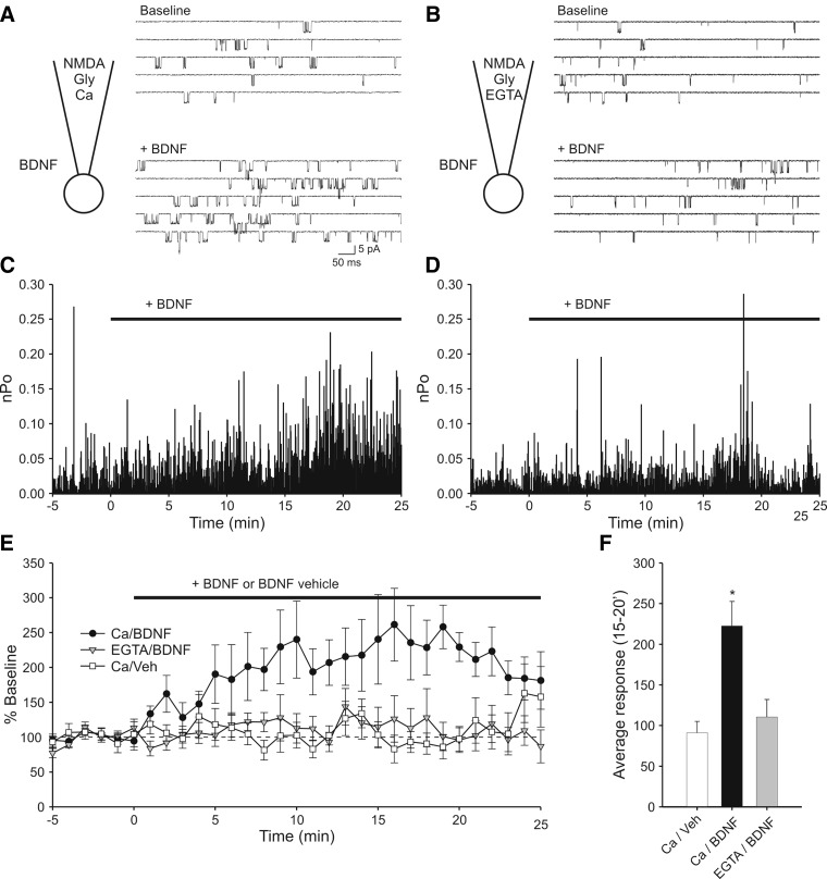
Brain-derived neurotrophic factor (BDNF), a potent modulator of synaptic transmission, is known to influence associative synaptic plasticity and refinement of neural connectivity. We now show that BDNF modulation of glutamate currents in hippocampal neurons exhibits the additional property of use dependence, a postsynaptic mechanism resulting in selective modulation of active channels. We demonstrate selectivity by varying the repetition rate of iontophoretically applied glutamate pulses during BDNF exposure. During relatively high-frequency glutamate pulses (0.1 Hz), BDNF application elicited a doubling of the glutamate current. During low-frequency pulses (0.0033 Hz), however, BDNF evoked a dramatically diminished response. This effect was apparently mediated by calcium because manipulations that prevented elevation of intracellular calcium largely eliminated the action of BDNF on glutamate currents. To confirm N-methyl-D-aspartate (NMDA) receptor involvement and assess spatial requirements, we made cell-attached single-channel recordings from somatic NMDA receptors. Inclusion of calcium in the pipette was sufficient to produce enhancement of channel activity by BDNF. Substitution of EGTA for calcium prevented BDNF effects. We conclude that BDNF modulation of postsynaptic NMDA receptors requires concurrent neuronal activity potentially conferring synaptic specificity on the neurotrophin's actions.


Figures

FIG. 1.
FIG. 1.
Brain-derived neurotrophic factor (BDNF)-induced potentiation of glutamate currents is activity-dependent. A: illustration of the recording configuration and stimulation protocols. Whole cell recordings (“patch” electrode) were made from the somata of cultured embryonic hippocampal neurons. Ionic current was elicited by iontophoretic application of glutamate to dendritic processes (“ionto” electrode) and BDNF or vehicle solution was bath-applied with a perfusion system (Ogata and Tatebayashi 1991). For the relatively high-frequency stimulation “activity” protocol, pulses of glutamate were delivered at 0.1 Hz for the duration of the recording. The diagram shows the 20-min period that begins at the onset of BDNF or vehicle perfusion. For the lower stimulation frequency “low activity” protocol, a single pulse of glutamate was delivered at 5-min intervals. After 20 min, stimulation frequency was increased to 0.1 Hz. B: example sweeps of glutamate currents during baseline period and 20 min into BDNF exposure. Repetitive glutamate application at 0.1 Hz (activity protocol) resulted in a twofold increase in total glutamate current (left). Reduction of stimulation frequency to 0.0033 Hz (low activity protocol) during BDNF exposure elicited a much smaller response (right). These and subsequent examples are the average of 3 sweeps from a single minute. C: averaged time courses of the response to BDNF during the activity (▪, n = 8) or low activity (⧫, n = 7) stimulation protocols. Peak current amplitude was normalized to baseline. Time = 0 has been set to onset of the BDNF perfusion. In this and subsequent figures, responses to 0.1-Hz stimuli were combined into 1-min bins and averaged. Responses to the 0.0033-Hz stimuli were not binned. —, the arithmetic addition of 2 time courses: BDNF during low activity, and activity alone from D (□ + ◊, see text). - - -, no response to BDNF. D: example sweeps of glutamate currents during baseline period and after 20 min of either the activity (left) or low activity (right) stimulation protocols. BDNF was not present during stimulation although cells were perfused with the BDNF vehicle solution in the same way as for BDNF. The activity protocol evoked a modest increase in glutamate current whereas the low activity protocol had no effect. E: averaged time courses of responses to activity (□, n = 10) and low activity protocols (◊, n = 7). Scale bars refer to sweeps in B and D.
FIG. 2.
FIG. 2.
Iontophoretic application of glutamate alone produces a modest increase in glutamate current via endogenously released BDNF. A: example sweeps of glutamate currents during baseline period and 20 min into the recording. Repetitive glutamate application at 0.1 Hz (activity protocol) produced an increase in glutamate current (left), which was prevented by bath application of K-252a (right). B: averaged time courses of 0.1-Hz stimulation alone + the K-252a/b vehicle (▪, n = 7) and 0.1-Hz stimulation in the presence of antagonists of neurotrophin signaling. K-252a (▿, n = 7), anti-BDNF function-blocking antibody (▵, n = 5), and TrkB-Fc (○, n = 6) reduced the increase in current seen in response to stimulation alone. The control compounds heat inactivated anti-BDNF antibody (▴, n = 7), and K-252b (▾, n = 7) had no effect. formula image 
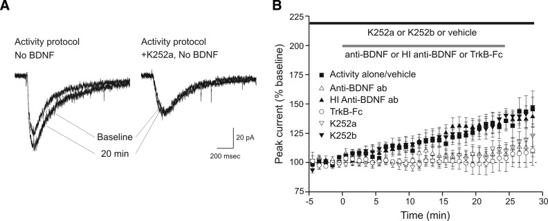
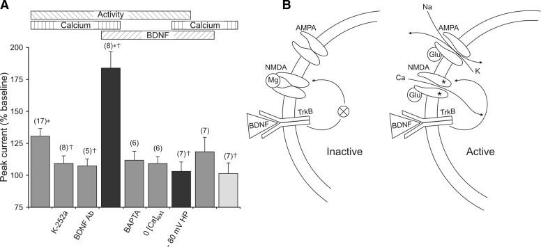
and □ (bars), application of the indicated compounds in separate sets of experiments. For all conditions, the activity protocol (0.1-Hz stimulation frequency) was used.
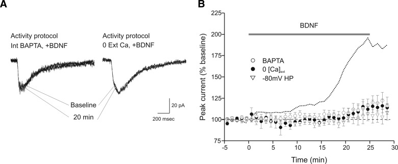
FIG. 3.
FIG. 3.
BDNF-induced potentiation of glutamate currents requires calcium influx and N-methyl-d-aspartate (NMDA) receptor activation. A: example sweeps of glutamate currents during baseline period and 20 min into BDNF exposure. BDNF application during repetitive glutamate application at 0.1 Hz (activity protocol) failed to produce a significant increase in glutamate current when 10 mM bis-(o-aminophenoxy)-N,N,N',N'-tetraacetic acid (BAPTA) was present in the recording pipette solution (left) or when calcium was not added to the bath solution (right). B: averaged time courses of responses to BDNF with 10 mM BAPTA included in the recording pipette solution (○, n = 6), bath solution nominally-free of calcium (•, n = 6), and cells held at –80 mV to prevent NMDA receptor openings (▿, n = 7). For comparison, a dotted line representing the time course of the normal response to BDNF during the activity protocol (from Fig. 1C) is included. For all conditions the activity protocol (0.1-Hz stimulation frequency) was used.
FIG. 4.
FIG. 4.
Glutamate receptor antagonist and change of holding potential during BDNF presentation confirms an absence of modulation when NMDA receptors are blocked. A: averaged time course of response to BDNF during co-application of kynurenate (n = 4 recordings). The holding potential was −40 mV, and the activity protocol was used. Block of the glutamate current during BDNF application prevented BDNF enhancement of current that was assessed following kynurenate washout. B: averaged time course of response to BDNF (n = 7 recordings). The holding potential was maintained at −80 mV except for the 2 indicated periods at −40 mV to assess the NMDA receptor component of current. As a result of changing the holding potential during these periods, the average amplitude of glutamate current increased slightly prior to BDNF perfusion and increased by a similar amount during BDNF perfusion. The data further support the conclusion that NMDA receptors are not being affected by BDNF in the absence of calcium influx.
FIG. 5.
FIG. 5.
Cell-attached single-channel recording shows that local calcium entry is sufficient for BDNF modulation of NMDA receptor activity. A: diagram illustrating the recording conditions and sample sweeps indicating modulation of NMDA receptor activity by BDNF. In addition to NMDA, glycine and other ingredients (see methods), the pipette solution contained 1.3 mM calcium. The patch contained ≥2 channels. Top: recording of baseline activity. Bottom: increased activity elicited by bath perfusion of 20 ng/ml BDNF. B: sample activity from a recording in which calcium in the pipette solution was replaced by EGTA. The patch contained ≥2 channels. Top and bottom traces: baseline activity and activity in the presence of BDNF, respectively. In this case, perfusion of BDNF had no effect on NMDA receptor activity. C: plot of open probability (Po) vs. time during for the recording illustrated in A. Time = 0 has been set to onset of the BDNF perfusion. Baseline activity is defined as the 5 min prior to BDNF perfusion. Note that Po increases with time during the BDNF perfusion. D: Po plot for the recording illustrated in B. Note that replacing Ca with EGTA in the pipette solution resulted in no effect of BDNF on NMDAR activity. E: plot in which Po values have been combined into 1-min bins and normalized to baseline activity. Experiments in which calcium was present in the pipette solution and which were exposed to BDNF show elevated activity (○, n = 10). Experiments in which the pipette contained calcium but were exposed to BDNF vehicle solution did not (□, n = 10). Experiments in which patches were exposed to BDNF but contained EGTA instead of calcium in the pipette solution also showed no increase in activity (▾, n = 9). F: summary graph in which average activity at 15–20 min during the BDNF perfusion is plotted. Addition of BDNF elicited a significant increase in activity as compared with vehicle when calcium was in the pipette solution (*, P < 0.01) but not when EGTA was in the pipette solution (P > 0.4).
FIG. 6.
FIG. 6.
Concurrence of activity, BDNF and extracellular calcium produced the largest enhancement of glutamate currents. A: horizontal bars (top) indicate the 3 stimulus conditions tested: 1) activity in the form of 0.1-Hz iontophoretic application of glutamate, 2) presence of extracellular calcium (or absence of augmented buffering of intracellular calcium), and 3) activation of TrkB by binding of BDNF. Vertical columns represent the magnitude of the increase in glutamate current observed in response to the different stimuli. The number of simultaneous conditions is represented by the color of the columns: 1 = light gray, 2 = dark gray, all 3 = black. Labels beneath the columns indicate special manipulations used to achieve the stimulus conditions. If no label is present, the condition was simply omitted. For example, the 1st 3 columns show results from experiments in which activity and calcium were present, but in which BDNF was not present or activation of TrkB by BDNF was prevented. This was accomplished by not adding BDNF (column 1), adding K-252a (column 2), or adding anti-BDNF (column 3). Overall, the figure shows that simultaneous use of activity, BDNF, and calcium produced the largest effect on glutamate current but only if NMDA receptors were allowed to open (compare columns 4 and 7). The number of experiments is noted at the top of each column. *, responses significantly different from calcium alone (column 9). †, responses significantly different from activity + calcium (column 1). B: model depicting BDNF modulation of glutamate current in active, but not inactive, cells. Left: BDNF binding to TrkB alone is not sufficient to elicit an effect on NMDA receptors because the cell and NMDA receptors are inactive. However, as shown on the right,synaptic release of glutamate produces a depolarization supplied by stimulation of AMPA receptors that also results in the opening of unblocked NMDA receptors. Consequently, calcium influx through NMDA receptors, together with BDNF binding to TrkB, results in enhancement of active NMDA receptor function.

Similar articles

Cited by

References

    1. Al-Hallaq RA, Conrads TP, Veenstra TD, Wenthold RJ. NMDA di-heteromeric receptor populations and associated proteins in rat hippocampus. J Neurosci 27: 8334–8343, 2007. - PMC - PubMed
    1. Amaral MD, Chapleau CA, Pozzo-Miller L. Transient receptor potential channels as novel effectors of brain-derived neurotrophic factor signaling: potential implications for Rett syndrome. Pharmacol Ther 113: 394–409, 2007. - PMC - PubMed
    1. Arvanian VL, Mendell LM. Acute modulation of synaptic transmission to motoneurons by BDNF in the neonatal rat spinal cord. Eur J Neurosci 14: 1800–1808, 2001. - PubMed
    1. Arvanov VL, Seebach BS, Mendell LM. NT-3 Evokes an LTP-like facilitation of AMPA/Kainate receptor-mediated synaptic transmission in the neonatal rat spinal cord. J Neurophysiol 84: 752–758, 2000. - PubMed
    1. Balkowiec A, Katz DM. Activity-dependent release of endogenous brain-derived neurotrophic factor from primary sensory neurons detected by ELISA in situ. J Neurosci 20: 7417–7423, 2000. - PMC - PubMed

Publication types

MeSH terms

LinkOut - more resources