. 2009 Jan;19(1):38-51.
doi: 10.1093/glycob/cwn102. Epub 2008 Oct 8.

N-Terminal clustering of the O-glycosylation sites in the Mycobacterium tuberculosis lipoprotein SodC

Affiliations

N-Terminal clustering of the O-glycosylation sites in the Mycobacterium tuberculosis lipoprotein SodC

Mark J Sartain et al. Glycobiology. 2009 Jan.

Abstract

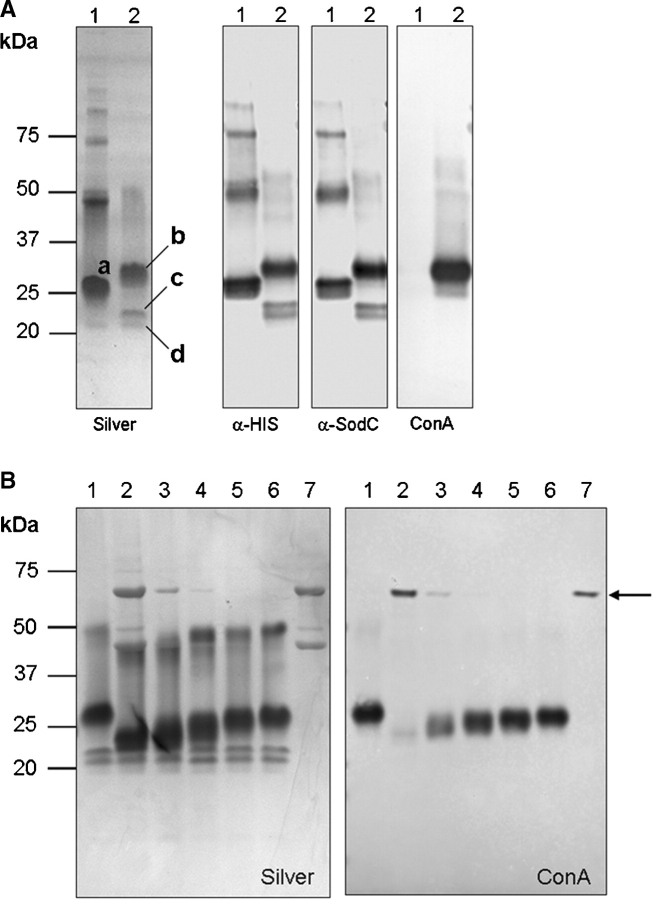
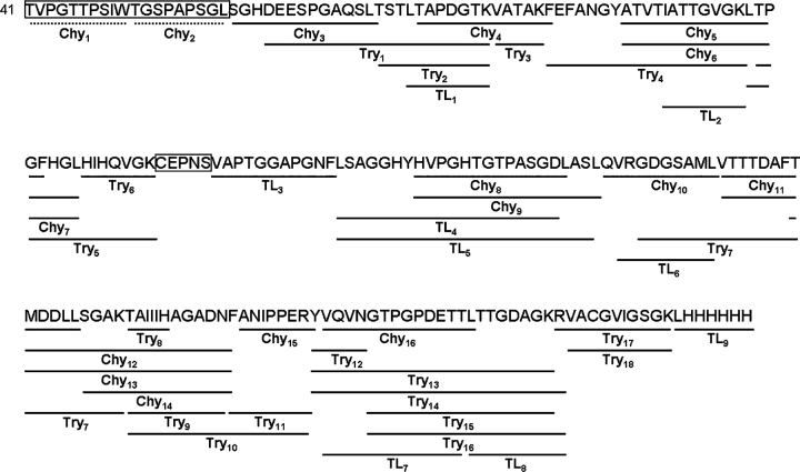
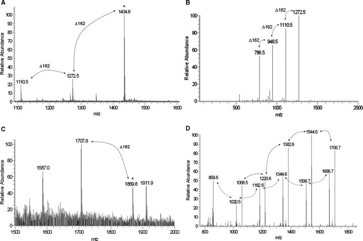
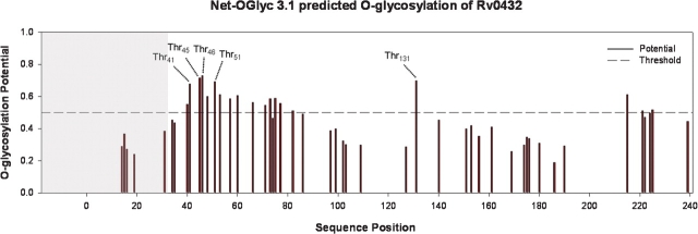
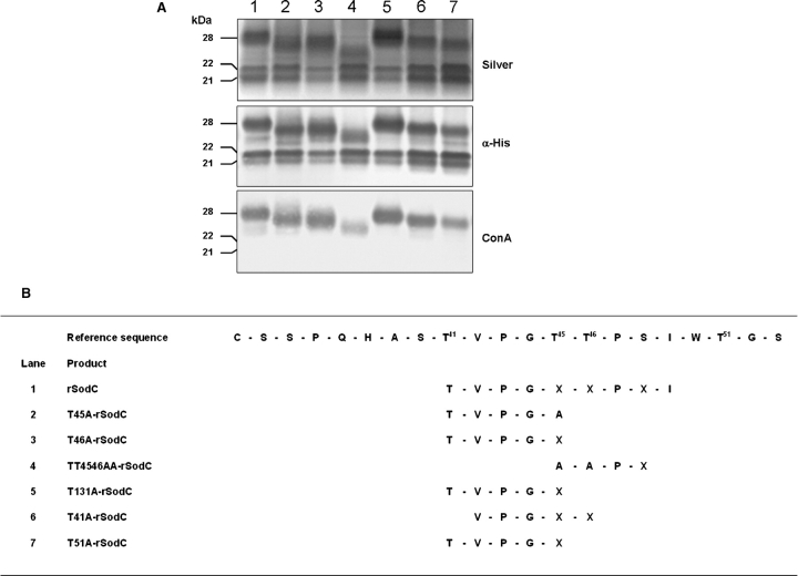
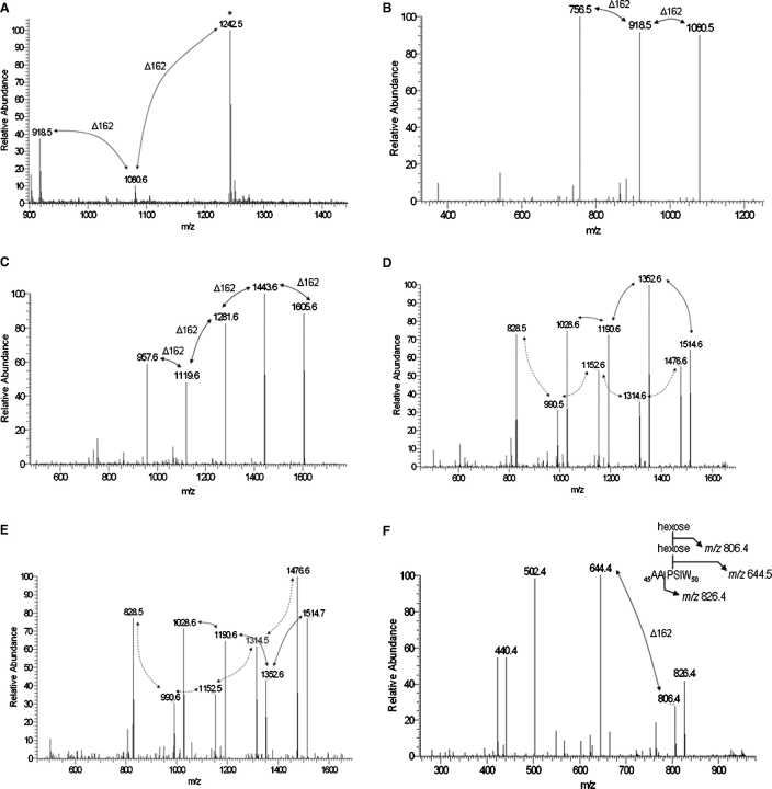
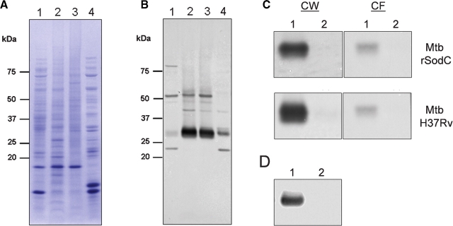
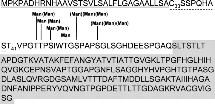
SodC is one of two superoxide dismutases produced by Mycobacterium tuberculosis. This protein was previously shown to contribute to virulence and to act as a B-cell antigen. SodC is also a putative lipoprotein, and like other Sec-translocated mycobacterial proteins it was suggested to be modified with glycosyl units. To definitively define the glycosylation of SodC, we applied an approach that combined site-directed mutagenesis, lectin binding, and mass spectrometry. This resulted in identification of six O-glycosylated residues within a 13-amino-acid region near the N-terminus. Each residue was modified with one to three hexose units, and the most dominant SodC glycoform was modified with nine hexose units. In addition to O-glycosylation of threonine residues, this study provides the first evidence of serine O-glycosylation in mycobacteria. When combined with bioinformatic analyses, the clustering of O-glycosylation appeared to occur in a region of SodC with a disordered structure and not in regions important to the enzymatic activity of SodC. The use of recombinant amino acid substitutions to alter glycosylation sites provided further evidence that glycosylation influences proteolytic processing and ultimately positioning of cell wall proteins.


Figures

Fig. 1
Fig. 1
ConA reactivity demonstrates glycosylation of Mtb rSodC. (A) Purified rSP-SodC (lane 1) and purified rSodC (lane 2) analyzed by SDS–PAGE and silver staining (panel 1) and Western blot analyses with anti-His5 monoclonal antibody (panel 2), polyclonal SodC antiserum (panel 3), and ConA as probes (panel 4). The proteins displayed are (a) the major 26-kDa product from E. coli; (b) the major 28-kDa product from Mtb; and (c and d) the minor 22- and 21-kDa products from Mtb. (B) Silver-stained SDS–PAGE (panel 1) and ConA Western blot (panel 2) analyses of purified rSodC from Mtb treated with various amounts of α-mannosidase. Lane 1, untreated rSodC; lanes 2–6, rSodC (10 μg) treated with 10, 2, 0.2, 0.1, and 0.02 μg α-mannosidase; and lane 7, α-mannosidase alone. The 44- and 66-kDa subunits of α-mannosidase are observed in the silver-stained gel (panel 1), and the 66-kDa subunit modified with a high-mannose-type-glycan (Kimura et al. 1999) displays ConA reactivity (denoted by arrow).
Fig. 2
Fig. 2
The deduced amino acid sequence (beginning with Thr41) of the 28-kDa rSodC protein product purified from Mtb and alignment with the peptides generated by chymotrypsin (Chy), trypsin (Try), and thermolysin (TL) digestion. The solid lines indicate the location of individual unmodified peptides identified by LC-ESI-MS/MS, and the boxes indicate unidentified sequences. The dashed lines indicate two predicted chymotrypsin-derived peptides used for the remainder of this work.
Fig. 3
Fig. 3
MS and MS/MS of glycopeptides generated by chymotrypsin digestion of rSodC. (A) An ESI-MS spectrum averaged over 80 scans corresponding to the Chy2 (51TGSPAPSGL59) peptide demonstrates primarily glycosylated forms modified with two, three, and four hexoses. (B) The m/z 1434.6 [M+H]+1 molecular ion marked by an asterisk in (A) was selected for ESI-MS/MS resulting in a fragmentation pattern containing neutral losses of 162 amu. The m/z 786.5 molecular ion represents the fully deglycosylated peptide. (C) An ESI-MS spectrum averaged over 96 scans corresponding to the Chy1 (41TVPGTTPSIW50) peptide demonstrates primarily glycosylated forms modified with four and five hexoses. (D) The m/z 1869.8 [M+H]+1 molecular ion marked by an asterisk in (C) was selected for ESI-MS/MS resulting in a fragmentation pattern containing neutral losses of 162 amu (marked with solid arrows). The m/z 1058.5 molecular ion represents the fully deglycosylated peptide. In addition, an ion series corresponding to a glycosylated y8 ion series with differences of 162 amu was observed (marked with dashed arrows).
Fig. 4
Fig. 4
Prediction of O-glycosylation sites for the complete Rv0432 protein sequence using the NetOglyc 3.1 algorithm (Julenius et al. 2005). The shaded region indicates the signal peptide. The labeled Thr residues indicate the predicted glycosylation sites targeted for amino acid substitution with Ala.
Fig. 5
Fig. 5
Analyses of purified rSodC proteins possessing Thr to Ala substitutions. (A) Silver-stained SDS–PAGE (top panel) and Western blot analyses with anti-His5 monoclonal antibody (middle panel) and ConA (bottom panel) as probes. Lane 1, nonmutated rSodC; lane 2, T45A-rSodC; lane 3, T46A-rSodC; lane 4, TT4546AA-rSodC; lane 5, T131A-rSodC; lane 6, T41A-rSodC; and lane 7, T51A-rSodC. (B) Amino-terminal sequencing of purified rSodC products. An “X” denotes the inability to assign an amino acid. The reference sequence provided begins with the putative lipidated Cys33 residue following signal peptide cleavage. Only the high-mass (26- to 28-kDa) products were sequenced.
Fig. 6
Fig. 6
MS and MS/MS of glycopeptides generated by chymotrypsin digestion of rSodC products with Thr to Ala substitutions. (A) An MS-ESI spectrum averaged over 89 scans corresponding to Chy2 (51AGSPAPSGL59) peptide of T51A-rSodC demonstrates primarily glycosylated forms modified with one to three hexoses. (B) The m/z 1242.5 [M+H]+1 molecular ion marked by an asterisk in (A) was selected for ESI-MS/MS resulting in a fragmentation pattern containing neutral losses of 162 amu. The m/z 756.5 molecular ion represents the fully deglycosylated peptide. (C) The m/z 1768.8 [M+H]+1 molecular ion corresponding to the Chy1 (42VPGTTPSIW50) peptide of T41A-rSodC with five hexoses was selected for ESI-MS/MS resulting in a fragmentation pattern containing neutral losses of 162 amu. The m/z 957.6 molecular ion represents the fully deglycosylated peptide. (D) The m/z 1677.7 [M+H]+1 molecular ion corresponding to the Chy1 (41TVPGATPSIW50) peptide of T45A-rSodC with four hexoses was selected for ESI-MS/MS resulting in a fragmentation pattern containing neutral losses of 162 amu (marked with solid arrows). The m/z 1028.6 molecular ion represents the fully deglycosylated peptide. In addition, an ion series corresponding to a glycosylated y8 ion series with differences of 162 amu was observed (marked with dashed arrows). (E) The m/z 1677.7 [M+H]+1 molecular ion corresponding to the Chy1 (41TVPGTAPSIW50) peptide of T46A-rSodC with four hexoses was selected for ESI-MS/MS resulting in a fragmentation pattern containing neutral losses of 162 amu (marked with solid arrows). The m/z 1028.6 molecular ion represents the fully deglycosylated peptide. In addition, an ion series corresponding to a glycosylated y8 ion series with differences of 162 amu was observed (marked with dashed arrows). (F) The m/z 969.0 [M+H]+1 molecular ion corresponding to the Chy1 (45AAPSIW50) peptide of TT4546AA-rSodC with two hexoses was selected for ESI-MS/MS resulting in a fragmentation pattern containing neutral losses of 162 amu. The m/z 644.4 molecular ion represents the fully deglycosylated peptide. The inset shows the fragmentation of this glycopeptide.
Fig. 7
Fig. 7
SodC subcellular localization. Subcellular fractions of Mtb H37Rv (lane 1, Cyt; lane 2, Mem; lane 3, CW; and lane 4, CF) were separated by SDS–PAGE and stained with Coomassie brilliant blue (A) or electroblotted and probed with polyclonal SodC antiserum (B). (C) Western blot with polyclonal SodC antiserum of TX-114 detergent phase partitioning (lane 1, detergent phase; lane 2, aqueous phase) performed on CW (left panels) and CF (right panels) fractions of Mtb expressing rSodC (top panels) and wild type Mtb H37Rv (bottom panels). (D) Western blot with polyclonal SodC antiserum of TX-114 detergent phase partitioning (lane 1, detergent phase; lane 2, aqueous phase) performed on WCL of M. smegmatis SP-rSodC.
Fig. 8
Fig. 8
Working model for the posttranslational modification of Mtb SodC. The region underlined with a solid line denotes the Type II signal peptide preceding the putative N-terminal acylated Cys (C33) described in D’Orazio et al. (2001). The region underlined with the dashed line denotes the experimentally determined truncation that resulted in an N-terminal Thr (T41). Glycosylation sites modified with mannose (Man) are indicated. Parentheses denote variable levels of glycosylation. The shaded region indicates the folded enzymatic structure, for which a more detailed annotation can be found in Figure S1 and Spagnolo et al. (2004).

Similar articles

Cited by

References

    1. Abernathy JL, Wang Y, Eckhardt AE, Hill RL. Identification of O-glycosylation sites with a gas phase sequencer. In: Abernathy JL, editor. Techniques in Protein Chemistry. New York: Academic Press; 1992. pp. 277–286.
    1. Battistoni A, Rotilio G. Isolation of an active and heat-stable monomeric form of Cu, Zn superoxide dismutase from the periplasmic space of Escherichia coli. FEBS Lett. 1995;374:199–202. - PubMed
    1. Baumgartner M, Karst U, Gerstel B, Loessner M, Wehland J, Jansch L. Inactivation of Lgt allows systematic characterization of lipoproteins from Listeria monocytogenes. J Bacteriol. 2007;189:313–324. - PMC - PubMed
    1. Belisle JT, Braunstein M, Rosenkrands I, Andersen P. The proteome of Mycobacterium tuberculosis. In: Cole ST, editor. Tuberculosis and the Tubercle Bacillus. Washington, DC: ASM Press; 2005. pp. 235–260.
    1. Coligan JE. Current Protocols in Protein Science. 2001. p. 10.15.11. - PubMed

Publication types