. 2008 Dec;19(12):5360-72.
doi: 10.1091/mbc.e08-01-0080. Epub 2008 Oct 8.

Beclin 1 forms two distinct phosphatidylinositol 3-kinase complexes with mammalian Atg14 and UVRAG

Affiliations

Beclin 1 forms two distinct phosphatidylinositol 3-kinase complexes with mammalian Atg14 and UVRAG

Eisuke Itakura et al. Mol Biol Cell. 2008 Dec.

Abstract

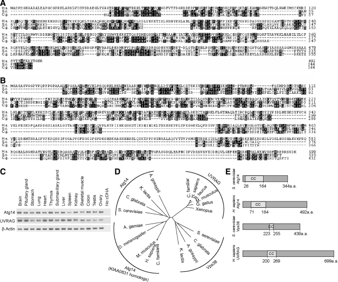
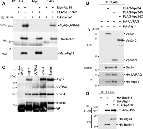
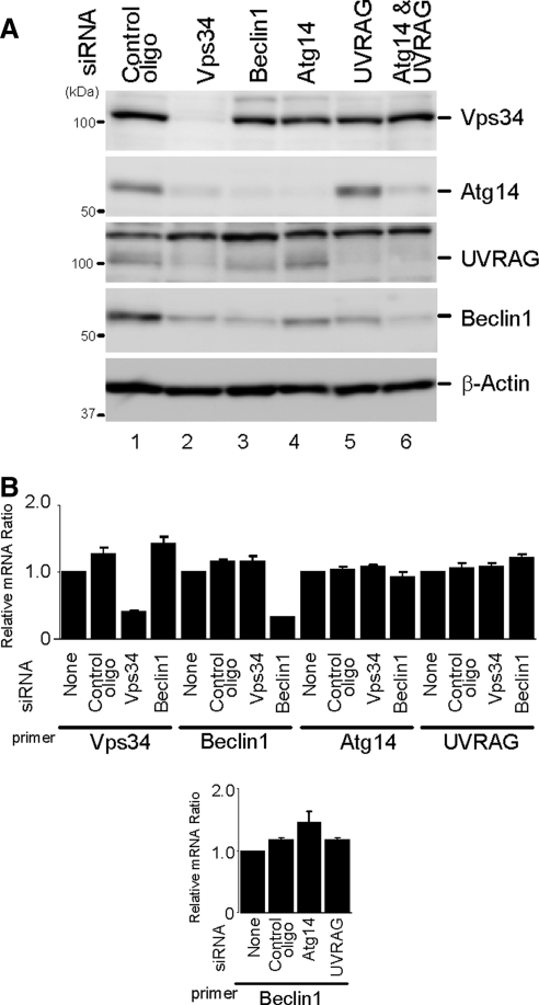
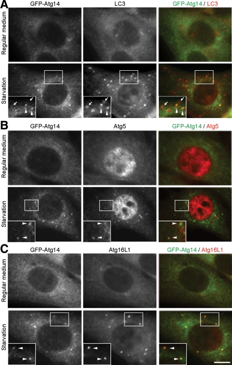
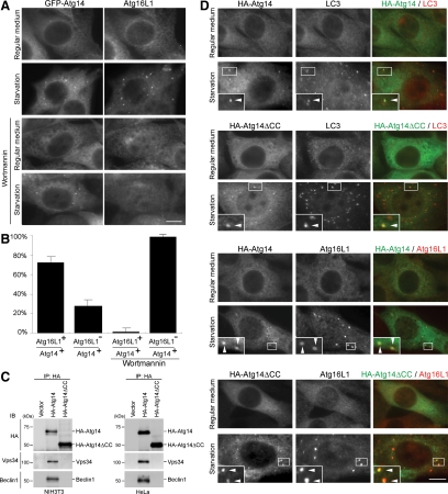
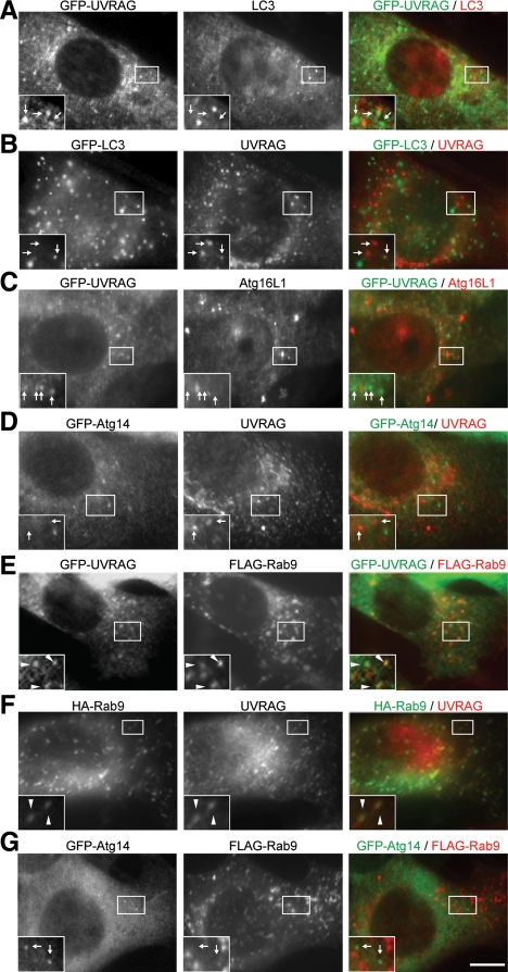
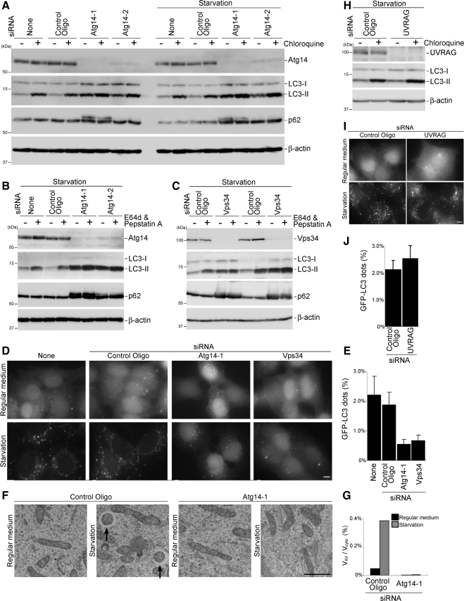
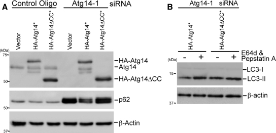
Class III phosphatidylinositol 3-kinase (PI3-kinase) regulates multiple membrane trafficking. In yeast, two distinct PI3-kinase complexes are known: complex I (Vps34, Vps15, Vps30/Atg6, and Atg14) is involved in autophagy, and complex II (Vps34, Vps15, Vps30/Atg6, and Vps38) functions in the vacuolar protein sorting pathway. Atg14 and Vps38 are important in inducing both complexes to exert distinct functions. In mammals, the counterparts of Vps34, Vps15, and Vps30/Atg6 have been identified as Vps34, p150, and Beclin 1, respectively. However, orthologues of Atg14 and Vps38 remain unknown. We identified putative mammalian homologues of Atg14 and Vps38. The Vps38 candidate is identical to UV irradiation resistance-associated gene (UVRAG), which has been reported as a Beclin 1-interacting protein. Although both human Atg14 and UVRAG interact with Beclin 1 and Vps34, Atg14, and UVRAG are not present in the same complex. Although Atg14 is present on autophagic isolation membranes, UVRAG primarily associates with Rab9-positive endosomes. Silencing of human Atg14 in HeLa cells suppresses autophagosome formation. The coiled-coil region of Atg14 required for binding with Vps34 and Beclin 1 is essential for autophagy. These results suggest that mammalian cells have at least two distinct class III PI3-kinase complexes, which may function in different membrane trafficking pathways.


Figures

Figure 1.
Figure 1.
Identification of mammalian counterparts of Atg14 and Vps38. (A) Amino acid alignment of Homo sapiens Atg14 (KIAA0831; National Center for Biotechnology Information [NCBI]accession no. NP_055739) with Atg14 of S. cerevisiae (accession no. NP_009686) and C. glabrata (accession no. XP_445209). The alignment was generated using CLUSTAL W. Identical residues are indicated with filled boxes. (B) Amino acid alignment of H. sapiens UVRAG (NCBI accession no. NP_003360) with Vps38 of S. cerevisiae (accession no. NP_013464) and C. glabrata (accession no. XP_445209). (C) KIAA0831 (Atg14) and UVRAG are ubiquitously expressed in mouse tissues. Total RNA from mouse tissues was reverse-transcribed into cDNA and then subjected to PCR amplification with indicated primers. (D) Phylogenetic analysis of Atg14, KIAA0831, UVRAG, and Vps38 homologues. The unrooted phylogenetic tree was constructed using CLUSTAL W based on the amino acid sequences of these homologues. Distance matrix-based trees were constructed by the NJ method (Saitou and Nei, 1987). (E) Structural comparison of S. cerevisiae Atg14, H. sapiens Atg14 (KIAA0831), S. cerevisiae Vps38, and H. sapiens UVRAG. The coiled-coil domain is predicted by the algorithm of Lupas et al. (1991).
Figure 2.
Figure 2.
Atg14 and UVRAG form two distinct complexes with Beclin 1-Vps34. (A) Human Atg14 and UVRAG are included in different Beclin 1 complexes. HEK293T cells were cotransfected with FLAG-UVRAG, HA-Beclin 1, and Myc-Atg14, and cell lysates were subjected to immunoprecipitation using antibodies against HA, Myc, and FLAG. The resulting precipitates were examined by immunoblot analysis with the indicated antibodies. (B) Atg14 and UVRAG interacts with the N-terminal C2 domain of Vps34. HEK293T cells were cotransfected with HA-UVRAG, HA-Atg14, and either FLAG-Vps34 (full-length), FLAG-Vps34N (containing C2 domain, 1-282 amino acid residues), or FLAG-Vps34C (283-887 amino acid residues) and analyzed as described in A. (C) Endogenous Atg14 and UVRAG form protein complexes with Vps34 and Beclin 1 but not with each other. Lysates of HEK293T cells were used for immunoprecipitation with indicated antiserum. (D) Atg14 forms a protein complex with p150. Lysates of HEK293T cells transiently expressing FLAG-p150 and either HA-Beclin 1 or HA-Atg14 were analyzed by immunoprecipitation and immunoblotting.
Figure 3.
Figure 3.
Beclin 1 and Vps34 are important for expression levels of Atg14 and UVRAG. (A) HeLa cells were treated with the indicated siRNA. Total cell lysates were prepared and analyzed by immunoblotting using the indicated antibodies. (B) Effect of each siRNA on mRNA expression levels. HeLa cells were treated with the indicated siRNAs for 72 h, and mRNA level was measured by real-time PCR. Data are expressed as mean ± SE of six PCR reactions.
Figure 4.
Figure 4.
Atg14 localizes on isolation membranes. NIH3T3 cells stably expressing GFP-Atg14 were cultured in regular DMEM or amino acid-free DMEM without FBS for 2 h. Then, cells were fixed, permeabilized, and subjected to immunofluorescence microscopy by using rat anti-GFP antibody and either polyclonal anti-LC3 (A), anti-Atg5 (B), or anti-Atg16L1 (C) antibody. Structures positive for both Atg14 and LC3 are indicated by arrowheads, and structures positive for LC3 but negative for Atg14 are indicated with arrows. Bar, 10 μm.
Figure 5.
Figure 5.
Atg14 puncta formation is independent of PI3-kinase activity. (A and B) Wortmannin inhibits puncta formation of Atg16L1 but not that of Atg14. NIH3T3 cells stably expressing GFP-Atg14 were cultured in regular DMEM or amino acid-free DMEM without FBS in the presence or absence of 200 nM wortmannin for 1 h. Then, cells were fixed, permeabilized, and subjected to immunofluorescence microscopy by using rat anti-GFP antibody and anti-rabbit Atg16L1 antibody. Bar, 10 μm (A). Atg16L1-positive rate of the Atg14 puncta under starvation conditions with or without wortmannin is shown as a percentage (B). Data are expressed as mean ± SD of at least 10 cells. (C) Atg14 interacts with Vps34 and Beclin 1 via the coiled-coil domain. Lysates were prepared from NIH3T3 cells stably expressing HA-Atg14 or HA-Atg14ΔCC and from HeLa cells transiently expressing HA-Atg14 or HA-Atg14ΔCC, and then they were subjected to immunoprecipitation analysis using anti-HA antibody. (D) Atg14 localizes on isolation membranes independently of binding to Vps34. NIH3T3 cells stably expressing HA-Atg14 or HA-Atg14ΔCC were cultured in regular DMEM or amino acid-free DMEM without FBS for 1 h. Then, cells were fixed, permeabilized, and subjected to immunofluorescence microscopy by using mouse anti-HA antibody and either polyclonal anti-LC3 or anti-Atg16L1 antibody. Structures positive for both markers are indicated by arrowheads. Bar, 10 μm.
Figure 6.
Figure 6.
UVRAG localizes on Rab9-positive late endosomes. NIH3T3 cells (F) and NIH3T3 cells stably expressing either GFP-UVRAG (A, C, and E), GFP-LC3 (B), or GFP-Atg14 (D and G) were transiently transfected with FLAG-Rab9 (E and G), or HA-Rab9 (F) for 24 h. In the experiments in B, C, D, and G, cells were subjected to starvation by culturing in amino acid-free DMEM without FBS for 2 h before fixation. After fixation, the cells were subjected to immunocytochemistry by using rat anti-GFP (A–E and G), rabbit anti-LC3 (A), rabbit anti-Atg16L1 (C), rabbit anti-FLAG (E and G), mouse anti-HA (F), or rabbit anti-UVRAG (B, D and F) antibodies. Structures positive for both green and red signals are indicated by arrowheads, and those positive for either a green or red signal are indicated with arrows. Bar, 10 μm.
Figure 7.
Figure 7.
Vps34 localizes on UVRAG-positive puncta and starvation-induced Atg14-positive puncta. NIH3T3 cells stably expressing Vps34-GFP and either HA-UVRAG (A) or HA-Atg14 (B) were generated. (B) Cells were subjected to starvation by culturing in amino acid-free DMEM without FBS for 2 h. After fixation, the cells were subjected to immunocytochemistry using rat anti-GFP and mouse anti-HA antibodies. Structures positive for both green and red signals are indicated by arrowheads. Bar, 10 μm.
Figure 8.
Figure 8.
Atg14 and Vps34 are required for autophagosome formation. (A–C) Reduced autophagic flux in cells treated with Atg14 (A and B) and Vps34 siRNA (C). HeLa cells treated with the indicated siRNA were incubated in regular DMEM or starvation medium in the presence or absence of chloroquine (20 μM) for 1 h (A) or E64d (50 μM) and pepstatin A (50 μg/ml) for 2 h (B and C). Total lysates of the cells were analyzed by immunoblotting by using the indicated antibodies. (D and E) HeLa cells stably expressing GFP-LC3 were treated with Atg14 and Vps34 siRNA as described in A–C and starved for 2 h. Bar, 10 μm (D). The ratio of the total area of GFP-LC3 dots to the total cellular area in starved cells is shown as a percentage. Data are expressed as mean ± SD of at least 10 different fields (E). (F and G) HeLa cells treated with the control or Atg14 siRNA were starved for 2 h and subjected to conventional electron microscopic analysis. Autophagosomes are indicated by arrows. Bar, 1 μm (F). The ratio of total autophagosome area to total cytoplasmic area was determined by morphometric analysis. Ten cells were analyzed for each sample using MetaMorph image analysis software (G). (H) HeLa cells treated with UVRAG siRNA were incubated in starvation medium in the presence or absence of chloroquine (20 μM) for 1 h. (I and J) HeLa cells stably expressing GFP-LC3, which were treated with UVRAG siRNA, were starved for 2 h and GFP-LC3 puncta were observed. Bar, 10 μm (I). The ratio of the total area of GFP-LC3 dots to the total cellular area in starved cells is shown as a percentage. Data are expressed as mean ± SD of at least 10 different fields (J).
Figure 9.
Figure 9.
Atg14ΔCC mutant cannot rescue the autophagy-defective phenotype of Atg14-knockdown cells. (A and B) HeLa cells were treated with the indicated siRNA. After 2 d, the cells were transiently transfected with an empty vector, HA-Atg14* (asterisk [*] indicates the siRNA-resistant version) or HA-Atg14ΔCC* plasmid together with the same siRNAs again, and then they were cultured for an additional 2 d. The cells were again transfected with the same expression vectors and cultured for another 1 d. Then, the cells were incubated in starvation medium for 2 h. Total lysates were prepared and p62 expression level (A) and LC3 turnover (B) were determined.

References

    1. Altschul S. F., Madden T. L., Schaffer A. A., Zhang J., Zhang Z., Miller W., Lipman D. J. Gapped BLAST and PSI-BLAST: a new generation of protein database search programs. Nucleic Acids Res. 1997;25:3389–3402. - PMC - PubMed
    1. Barbero P., Bittova L., Pfeffer S. R. Visualization of Rab9-mediated vesicle transport from endosomes to the trans-Golgi in living cells. J. Cell Biol. 2002;156:511–518. - PMC - PubMed
    1. Berg T. O., Fengsrud M., Stromhaug P. E., Berg T., Seglen P. O. Isolation and characterization of rat liver amphisomes. Evidence for fusion of autophagosomes with both early and late endosomes. J. Biol. Chem. 1998;273:21883–21892. - PubMed
    1. Bjørkøy G., Lamark T., Brech A., Outzen H., Perander M., Øvervatn A., Stenmark H., Johansen T. p62/SQSTM1 forms protein aggregates degraded by autophagy and has a protective effect on huntingtin-induced cell death. J. Cell Biol. 2005;171:603–614. - PMC - PubMed
    1. Blommaart E. F., Krause U., Schellens J. P., Vreeling-Sindelarova H., Meijer A. J. The phosphatidylinositol 3-kinase inhibitors wortmannin and LY294002 inhibit autophagy in isolated rat hepatocytes. Eur. J. Biochem. 1997;243:240–246. - PubMed

Publication types

MeSH terms