. 2008:14:1784-91.
Epub 2008 Oct 6.

Mature retinal pigment epithelium cells are retained in the cell cycle and proliferate in vivo

Affiliations

Mature retinal pigment epithelium cells are retained in the cell cycle and proliferate in vivo

Heba Al-Hussaini et al. Mol Vis. 2008.

Abstract

Purpose: To investigate the capacity of mature retinal pigment epithelium (RPE) cells to enter the cell cycle in vivo using a range of RPE-specific and proliferative specific markers in both pigmented and albino rats.

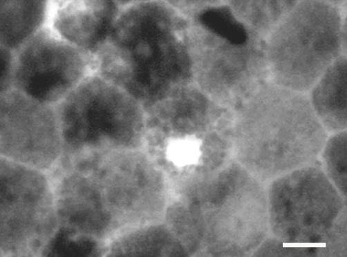
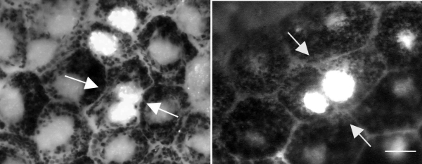
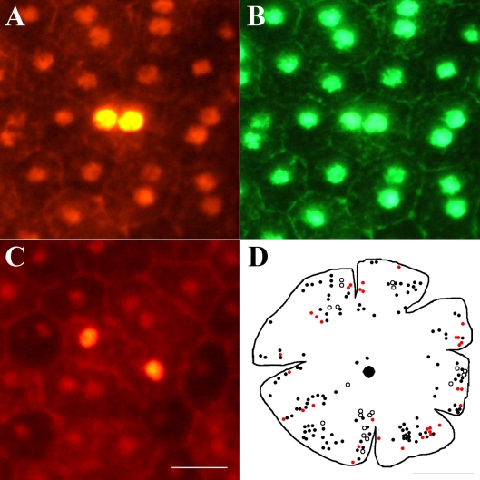
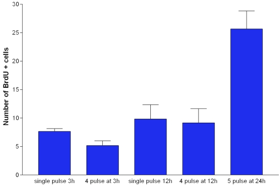
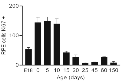
Methods: Whole-mounted retinas of both Dark Agouti and albino rats were immunolabeled with cell cycle markers Ki67 or PCNA and double labeled with RPE cell marker RPE65 or CRALBP. The number and distribution of these cells was mapped. An additional group of Dark Agouti rats were given repeated intraperitoneal injections of Bromodeoxyuridine (BrdU )for 20 days and then sacrificed 30 days later. The retinas were then processed for BrdU detection and Otx, a RPE cell-specific marker. For comparison, human RPE tissue from a postmortem donor was also labeled for Ki67.

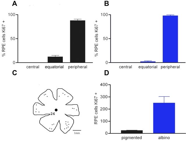
Results: In both pigmentation phenotypes, a subpopulation of mature RPE cells in the periphery were positive for both cell cycle markers. These cells were negative for Caspase 3, hence were not apoptotic. Ki67-positive cells were also seen in human RPE. Further, many cells positive for BrdU were identified in similar retinal regions, confirming that RPE cells not only enter the cell cycle but also divide, albeit at a slow cell cycle rate. There was a ten fold increase in the number of RPE cells positive for cell cycle markers in albino (approximately 200 cells) compared to pigmented rats (approximately 20 cells).

Conclusions: Peripheral RPE cells in rats have the capacity to enter the cell cycle and complete cellular division.


Figures

Figure 1
Figure 1
Labeling patterns in retinal pigment epithelium (RPE) sheets in whole mount preparations taken from DA rats. A: RPE cells positively labeled for Ki67. These appear to be in anaphase. B: This is the same region as shown in A, but stained with RPE65, which is an RPE specific marker in this tissue. C: This is the same region as shown in A and B but stained with DAPI to reveal the nuclei of the imaged cells. D-F: These are stained in the same way as A-C, however here the cell positive for Ki67 shown in D has down-regulated RPE65 as shown in E. F is the corresponding DAPI stained image. G: This shows a Ki67 positive RPE cell which has also in H been stained with CRALBP, which is a second RPE specific marker in this tissue. I is the same region stained with DAPI to reveal nuclei. J shows a Ki67 positive RPE cells. Taken in black and white the melanin granules in the cell can be clearly identified (arrow), which along with the RPE65 and CRALBP confirm that the tissue sheet examined is RPE. K shows an RPE cell positive for a second cell cell cycle marker PCNA, and L shows the same image stained with DAPI. The scale bar represents 10 µm.
Figure 2
Figure 2
The distribution and number of Ki67 positive RPE cells in the retinae of pigmented (black bars) and albino (blue bars) rats. A and B show the relative distribution of Ki67 cells in retinae of pigmented and albino rats. In both cases the largest percentage of the total number of cells are located in the periphery with none in central retina regions. The actual distribution of these cells in a pigmented rat is given in C. D shows the absolute number of Ki67 positive RPE cells in a pigmented and an albino animals. The differences shown in A and B between the different retinal regions were statistically significant (ANOVA p<0.0001). Differences between equatorial and peripheral regions were also statistically significant (Newman Keules p<0.001). While the relative differences in the distribution of Ki67 cells within retinae was similar between pigmentation phenotypes, there were always many more RPE cells labeled with Ki67 in albinos compared with pigmented rats. This difference was statistically significant (Newman-Keuls p<0.01).
Figure 3
Figure 3
Ki67-positive cells in the sample of human eye tissue. Only a small strip of tissue, spanning from the equatorial to peripheral regions, was examined. While Ki67-positive cells were clearly present, it was not possible to estimate the number of these cells or map their retinal location. Scale bar equals 10 µm.
Figure 4
Figure 4
The percentage of binucleated RPE cells in different retinal regions in pigmented (black bars) and albino (blue bars) rats. In adult rodent retinas, many of the RPE cells were binucleated. The proportion of these have been determined in both pigmented (A) and albino (B) retinas. The retinas were divided into three roughly equal geographic regions: central, equatorial, and peripheral. In both pigmentation phenotypes the majority of the binucleated cells were located toward the central retina, although many were also found in equatorial regions. The distribution of these cells is the reverse pattern of that found for Ki67-positive cells shown in Figure 2. The differences in A and B are statistically significant (ANOVA, p<0.001). In both cases, the differences between the percentage of binucleated RPE cells between central and equatorial regions were not statistically significant. However, differences in the percentage of binucleated RPE cells found central and peripheral regions were significantly different (Newman-Keuls p<0.01). The differences between equatorial and peripheral regions were also statistically significant (Newman-Keuls p<0.01).
Figure 5
Figure 5
RPE cells were identified that appear to be going through full cell division. In the peripheral retinal regions, a small number of cells could be identified that appeared to be passing through full cell division. In both pictures, arrows point to two labeled nuclei that appear to be forming a plasma membrane between them. Both sets of cells appear irregular in the RPE cell matrix. Taken together with the finding that there was no increase in the number of binucleated cells in the peripheral retina, these photographs demonstrate that at least some of the cells in this region were undergoing full cell division.
Figure 6
Figure 6
BrdU labeling in mature RPE cells. A and B: Shows a binucleated labeled cell with BrdU on red channel and Otx green channel. C shows two adjacent mono-nucleated cells that are labeled with BrdU. Scale bar equals 20 µm. D shows an outline drawing on which the location of RPE cells labeled with Ki67 and BrdU are marked. The diagram shows the distribution of positive BrdU-labeled binucleated (black dots) and mononucleated (black circle) cells. Only a small number of the BrdU-labeled cells were more centrally located than those labeled for Ki67. The mononucleated cells were almost always found in pairs of close proximity. The red dots represent the number and distribution of Ki67 positive RPE cells. While these largely overlap with the BrdU labeled population of RPE cells, they tend to occupy a slightly more peripheral location. The scale bar represents 2.5 mm.
Figure 7
Figure 7
The number of BrdU positive cells in the RPE following multiple pulses at progressive times. BrdU was given as a single pulse (50µg/kg) or as multiple pulses in 2 month old DA rats. For comparison animals were given 4 injections at 3 h intervals and euthanized 3 h later. Similar experiments were undertaken at 12 h intervals, with single pulse and euthanized 12 h later compared with 4 pulses separated by 12 h each and euthanized 12 h after the last pulse. There was no statistically significant increase in labeled cells following multiple injections compared with single injections. However, when BrdU was pulsed at 24 h intervals over 5 days, there was a significant increase in the number of RPE labeled cells compared with labeling in any of the other groups (ANOVA, p<0.0001. Dunnett’s Multiple Comparison Test p<0.001). Hence the RPE population is proliferating, but with a cell cycle rate of approximately 5 days.
Figure 8
Figure 8
The number of Ki67-positive cells found in the RPE flat mounts of pigmented animals sampled from embryonic day 18 (E18) through to postnatal day 150. Relatively few cells were in the cell cycle at E18, which is when the normal patterns of cell division in developing tissue end. However there was a marked increase (ANOVA, p<0.0001) in the number of these cells on the day of birth (0), which was statistically significant compared to E18 (Newman-Keuls, p<0.001). From P0 on, cell number gradually declined with age.

References

    1. Curcio CA, Sloan KR, Kalina RE, Hendrickson AE. Human photoreceptor topography. J Comp Neurol. 1990;292:497–523. - PubMed
    1. Cunea A, Jeffery G. The ageing photoreceptor. Vis Neurosci. 2007;24:151–5. - PubMed
    1. Gao H, Hollyfield JG. Aging of the human retina. Differential loss of neurons and retinal pigment epithelial cells. Invest Ophthalmol Vis Sci. 1992;33:1–17. - PubMed
    1. Balazsi AG, Rootman J, Drance SM, Schulzer M, Douglas GR. The effect of age on the nerve fiber population of the human optic nerve. Am J Ophthalmol. 1984;97:760–6. - PubMed
    1. Panda-Jonas S, Jonas JB, Jakobczyk-Zmija M. Retinal pigment epithelial cell count, distribution, and correlations in normal human eyes. Am J Ophthalmol. 1996;121:181–9. - PubMed

Publication types

LinkOut - more resources