Comparative Study
. 2008 Oct;7(5):706-16.
doi: 10.1111/j.1474-9726.2008.00418.x.

Mitochondrial iron accumulation with age and functional consequences

Affiliations
Comparative Study

Mitochondrial iron accumulation with age and functional consequences

Arnold Y Seo et al. Aging Cell. 2008 Oct.

Abstract

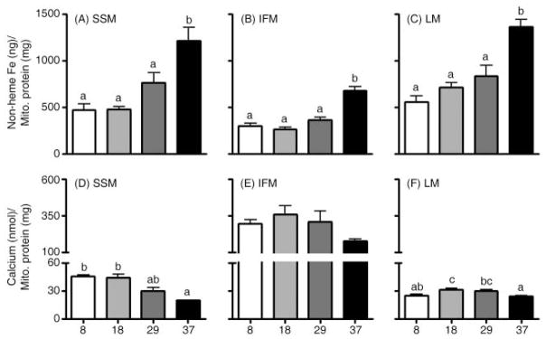
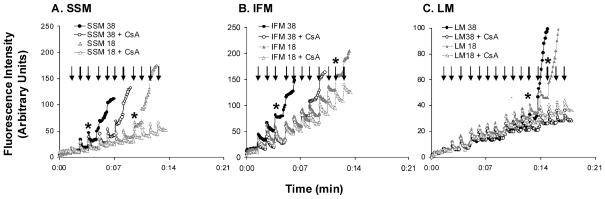
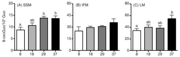
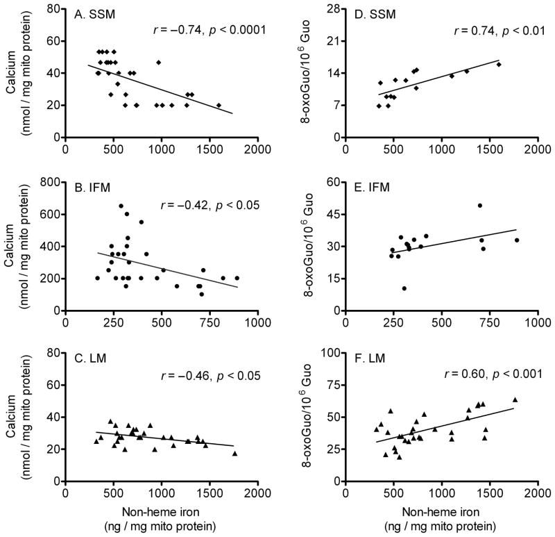
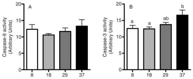
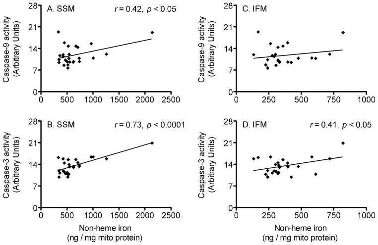
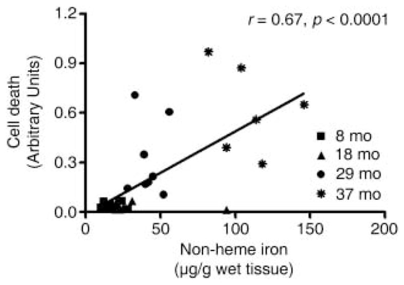
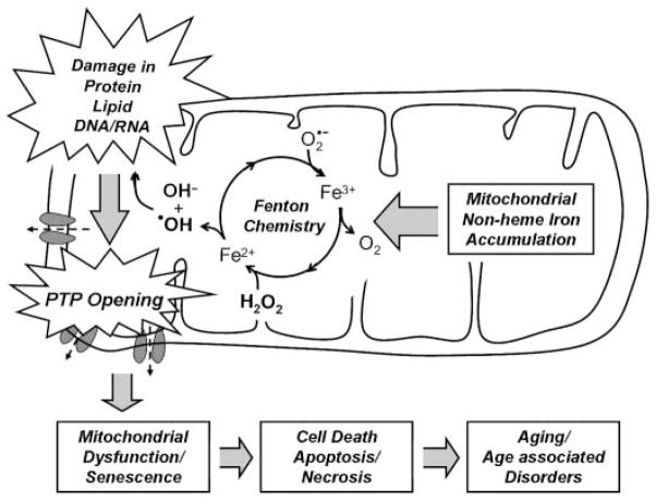
During the aging process, an accumulation of non-heme iron disrupts cellular homeostasis and contributes to the mitochondrial dysfunction typical of various neuromuscular degenerative diseases. Few studies have investigated the effects of iron accumulation on mitochondrial integrity and function in skeletal muscle and liver tissue. Thus, we isolated liver mitochondria (LM), as well as quadriceps-derived subsarcolemmal mitochondria (SSM) and interfibrillar mitochondria (IFM), from male Fischer 344 x Brown Norway rats at 8, 18, 29 and 37 months of age. Non-heme iron content in SSM, IFM and LM was significantly higher with age, reaching a maximum at 37 months of age. The mitochondrial permeability transition pore (mPTP) was more susceptible to the opening in aged mitochondria containing high levels of iron (i.e. SSM and LM) compared to IFM. Furthermore, mitochondrial RNA oxidation increased significantly with age in SSM and LM, but not in IFM. Levels of mitochondrial RNA oxidation in SSM and LM correlated positively with levels of mitochondrial iron, whereas a significant negative correlation was observed between the maximum Ca(2+) amounts needed to induce mPTP opening and iron contents in SSM, IFM and LM. Overall, our data suggest that age-dependent accumulation of mitochondrial iron may increase mitochondrial dysfunction and oxidative damage,thereby enhancing the susceptibility to apoptosis.


Figures

Fig. 1
Fig. 1
Effects of aging on mitochondrial non-heme iron levels and permeability transition pore opening (i.e. Ca2+-retention capacity) in skeletal muscle and liver. Non-heme iron levels increased with age in quadriceps (A) subsarcolemmal mitochondria (SSM), (B) interfibrillar mitochondria (IFM) and (C) liver mitochondria (LM) and showed the greatest accumulation after 29 months of age. Ca2+-retention capacity significantly decreased with age in (D) SSM, but not in (E) IFM (p = 0.068). (F) LM showed significantly decreased Ca2+-retention capacity in 37-month-old rats compared to 18-month-old rats. Data are expressed as mean ± SEM (n = 7–9). a,b,c Different letters indicate values are significantly different (p < 0.05).
Fig. 2
Fig. 2
Representative experiments of calcium-induced mitochondrial permeability transition pore (mPTP) opening. (A) subsarcolemmal mitochondria (SSM; 0.75 mg mL−1), (B) interfibrillar mitochondria (IFM; 0.1 mg mL−1) and (C) liver mitochondria (LM; 1.0 mg mL−1) were energized with glutamate/malate. 1.25 nmol (SSM and IFM) and 0.65 nmol (LM) of CaCl2 were added to mitochondria with a 1-min interval between injections. During this time, extra-mitochondrial Ca2+ pulses were recorded in the presence of 1 μM calcium green-5 N. Arrows indicate Ca2+ injections. Asterisks (*) denote the injections which open mPTP, leading to mitochondrial Ca2+ release. Incubation with 0.5 μM of cyclosporin A (CsA) could significantly inhibit mPTP opening.
Fig. 3
Fig. 3
Effects of aging on mitochondrial RNA oxidation in skeletal muscle and liver. Levels of mtRNA increased significantly with age in (A) subsarcolemmal mitochondria (SSM), whereas age did not affect RNA oxidation in (B) interfibrillar mitochondria (IFM). With age, RNA oxidation was significantly increased in (C) liver mitochondria. Data are expressed as mean ± SEM (n = 4–7). a,bDifferent letters indicate values are significantly different (p < 0.05).
Fig. 4
Fig. 4
Correlation between mitochondrial non-heme iron level, Ca2+-retention capacity and mtRNA oxidation. Correlation analyses were performed to determine how mitochondrial iron levels relate to mPTP opening and mtRNA oxidation. There was a significant negative correlation between iron levels and mPTP opening: (A) subsarcolemmal mitochondria (SSM; n = 32), (B) interfibrillar mitochondria (IFM; n = 29), and (C) liver mitochondria (LM; n = 30). In (D) SSM (n = 14), RNA oxidation levels was correlated with iron contents, but not in (E) IFM (p = 0.08, n = 16). A positive correlation between iron contents and RNA oxidation levels was detected in (F) LM (n = 30).
Fig. 5
Fig. 5
Cytosolic caspase-9 and caspase-3 activities. (A) Caspase-9 activity was not changed with age, whereas (B) caspase-3 activity was significantly increased in the aged rat quadriceps muscle (p < 0.05). Data are expressed as mean ± SEM (n = 4–8). a,bDifferent letters indicate values are significantly different (p < 0.05).
Fig. 6
Fig. 6
Correlation between mitochondrial non-heme iron level and caspase-9 and caspase-3 activities. Pearson’s tests were performed to determine whether mitochondrial iron levels correlate with caspase-9 and caspase-3 activities. Iron levels in subsarcolemmal mitochondria (SSM) significantly correlated with (A) caspase-9 activity (n = 26) and (B) caspase-3 activity (n = 26). There was no correlation between interfibrillar mitochondria’s (IFM) iron contents and (C) caspase-9 activity, whereas a positive correlation between iron levels and (D) caspase-3 activity was found in IFM (n = 25).
Fig. 7
Fig. 7
Correlation between non-heme iron level and cell death in rat gastrocnemius muscle at 8-, 18-, 29- and 37-month ages. Pearson’s tests were performed to determine whether non-heme iron levels correlate with muscle cell death. The correlation between non-heme iron and cell death was highly significant (n = 30).
Fig. 8
Fig. 8
Potential role of mitochondrial iron accumulation in the aging process. Accumulation of mitochondrial iron, possibly due to altered mechanisms of mitochondrial iron transport increases oxidative stress via Fenton chemistry and the decay of mitochondrial structural components, such as proteins, lipids and nucleic acids. With age, an increase in the susceptibility of the permeability transition pore (PTP) may cause mitochondrial dysfunction and cellular degeneration via apoptosis or necrosis.

References

    1. Adhihetty PJ, Ljubicic V, Menzies KJ, Hood DA. Differential susceptibility of subsarcolemmal and intermyofibrillar mitochondria to apoptotic stimuli. Am J Physiol Cell Physiol. 2005;289:C994–C1001. - PubMed
    1. Atamna H, Walter PB, Ames BN. The role of heme and iron-sulfur clusters in mitochondrial biogenesis, maintenance, and decay with age. Arch Biochem Biophys. 2002;397:345–353. - PubMed
    1. Babcock M, de SD, Oaks R, vis-Kaplan S, Jiralerspong S, Montermini L, Pandolfo M, Kaplan J. Regulation of mitochondrial iron accumulation by Yfh1p, a putative homolog of frataxin. Science. 1997;276:1709–1712. - PubMed
    1. Beinert H, Kiley PJ. Fe-S proteins in sensing and regulatory functions. Curr Opin Chem Biol. 1999;3:152–157. - PubMed
    1. Brunner EA, Cheng SC, Berman ML. Effects of anesthesia on intermediary metabolism. Annu Rev Med. 1975;26:391–401. - PubMed

Publication types

MeSH terms