. 2008 Oct;7(5):733-45.
doi: 10.1111/j.1474-9726.2008.00422.x.

p16INK4a-induced senescence is disabled by melanoma-associated mutations

Affiliations
Free PMC article

p16INK4a-induced senescence is disabled by melanoma-associated mutations

Sebastian Haferkamp et al. Aging Cell. 2008 Oct.
Free PMC article

Abstract

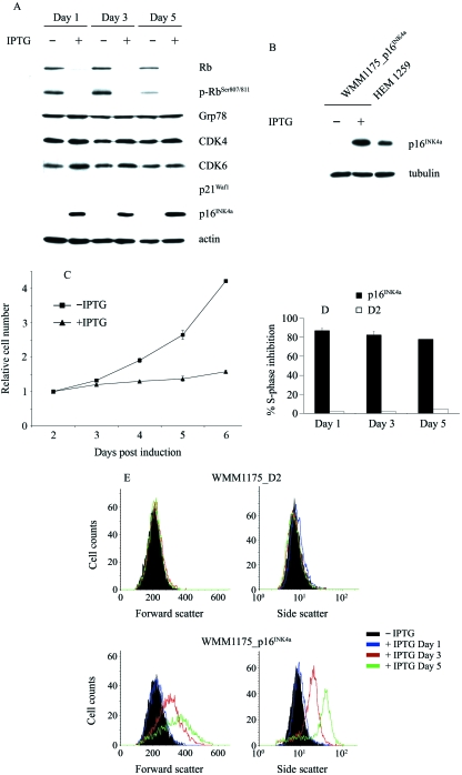
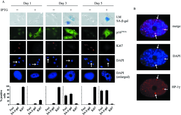
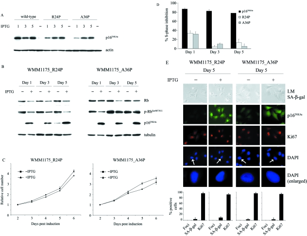
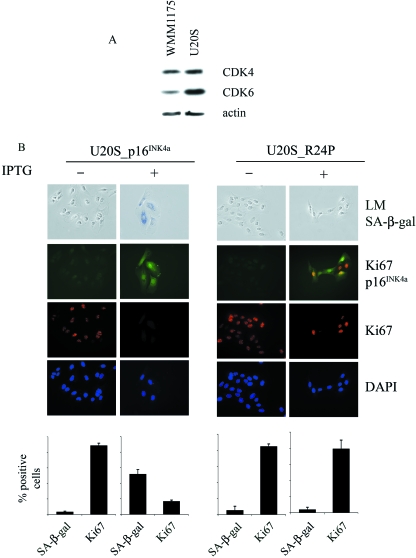
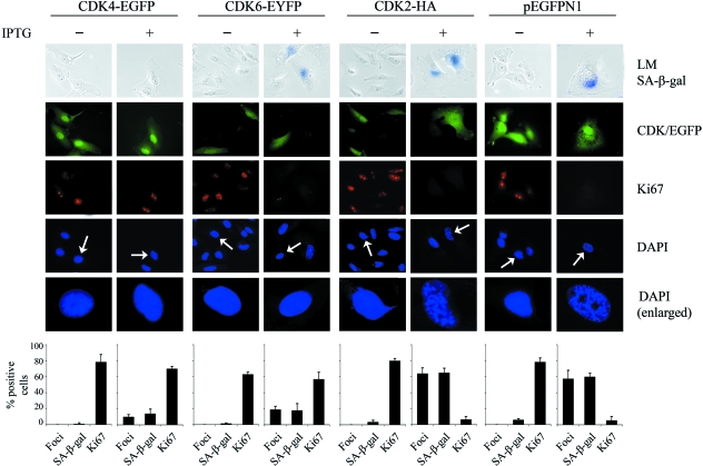
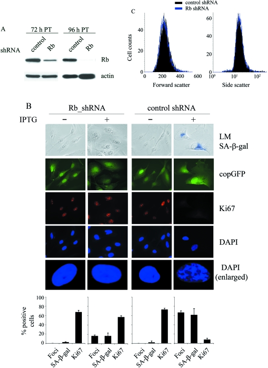
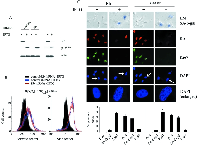
The p16(INK4a)-Rb tumour suppressor pathway is required for the initiation and maintenance of cellular senescence, a state of permanent growth arrest that acts as a natural barrier against cancer progression. Senescence can be overcome if the pathway is not fully engaged, and this may occur when p16(INK4a) is inactivated. p16(INK4a) is frequently altered in human cancer and germline mutations affecting p16(INK4a) have been linked to melanoma susceptibility. To characterize the functions of melanoma-associated p16(INK4a) mutations, in terms of promoting proliferative arrest and initiating senescence, we utilized an inducible expression system in a melanoma cell model. We show that wild-type p16(INK4a) promotes rapid cell cycle arrest that leads to a senescence programme characterized by the appearance of chromatin foci, activation of acidic beta-galactosidase activity, p53 independence and Rb dependence. Accumulation of wild-type p16(INK4a) also promoted cell enlargement and extensive vacuolization independent of Rb status. In contrast, the highly penetrant p16(INK4a) variants, R24P and A36P failed to arrest cell proliferation and did not initiate senescence. We also show that overexpression of CDK4, or its homologue CDK6, but not the downstream kinase, CDK2, inhibited the ability of wild-type p16(INK4a) to promote cell cycle arrest and senescence. Our data provide the first evidence that p16(INK4a) can initiate a CDK4/6-dependent autonomous senescence programme that is disabled by inherited melanoma-associated mutations.


Figures

Fig. 1
Fig. 1
Induced expression of p16INK4a inhibits Rb phosphorylation, limits cell proliferation and alters cell morphology. (A) Expression of the indicated proteins was determined by Western blot at 1, 3 and 5 days after treatment of WMM1175_p16INK4a cells with 4 mm IPTG. (B) Accumulation of p16INK4a in IPTG-treated (5 days) WMM1175_p16INK4a cells compared with levels of endogenous p16INK4a in normal, actively proliferating human neonatal epidermal melanocytes (HEM 1259). (C) The impact of induced p16INK4a expression on the proliferation of the WMM1175_p16INK4a cells was determined over a 5-day induction period using the MTS assay. The results shown are expressed as the average ± standard deviation of at least two independent experiments performed in triplicate. (D) The percentage of cells in S-phase after induction of p16INK4a for up to 5 days was determined by flow cytometry. S-phase inhibition was calculated from at least two independent induction experiments. Percentage S-phase inhibition in the IPTG-treated parental WMM1175_D2 cells, which expresses the lac repressor, but not the p16INK4a transgene, is also shown. (E) The impact of IPTG-exposure on the size (Forward scatter) and granularity (Side scatter) of the WMM1175_p16INK4a melanoma cells and the parental WMM1175_D2 cell line was investigated using flow cytometry on unfixed cells. These results are representative of at least two independent experiments.
Fig. 2
Fig. 2
Impact of induced p16INK4a expression on the cellular senescence programme. (A) WMM1175_p16INK4a cells were exposed to 4 mm IPTG over a 5-day period. The accumulation of p16INK4a, cell proliferation (Ki67), chromatin condensation (DAPI) and the appearance of SA-β-gal was analysed. Cells enlarged to show DAPI-stained chromatin foci are indicated with arrows. Cell counts for each of these markers are shown as histograms, which correspond to the average ± standard deviation of at least two independent induction experiments from a total of at least 500 cells. LM, light microscopy. (B) Representative examples of p16INK4a-induced chromatin condensation (DAPI) and costaining for HP-1γ as surrogates for senescence-associated heterochromatin foci (indicated by arrows).
Fig. 3
Fig. 3
Melanoma-associated p16INK4a mutants fail to induce cell cycle arrest. (A) Expression of wild-type p16INK4a, R24P and A36P mutant proteins in WMM1175 melanoma cell clones was induced with 4 mm IPTG over a 5-day induction period and compared using immunoblotting. (B) Expression of p16INK4a, total Rb, Ser807/811-phosphorylated Rb and actin was determined 1, 3 and 5 days after treatment of WMM1175_R24P and WMM1175_A36P cells with 4 mm IPTG. (C) The impact of induced mutant p16INK4a on the proliferation of the WMM1175_R24P and WMM1175_A36P cells was determined over the 5-day induction period using the MTS assay. The results shown are expressed as the average ± standard deviation of at least two independent experiments performed in triplicate. (D) The percentage of cells in S-phase after induction of p16INK4a or a melanoma-associated p16INK4a mutant in the WMM1175 cells was determined by flow cytometry. The percentage S-phase inhibition was calculated from at least two independent induction experiments. (E) Expression of the melanoma-associated variants R24P or A36P was induced in the WMM1175 melanoma cells and the impact on cell morphology (LM), proliferation (Ki67), chromatin condensation (DAPI) and SA-β-gal activity was analysed over a 5-day period. Representative examples of the 5-day IPTG induction point are shown. Cells enlarged to show DAPI-stained chromatin foci are indicated with arrows. Cell counts for the cell cycle markers are shown as histograms, which correspond to the average ± standard deviation of at least two independent induction experiments from a total of at least 500 cells. LM, light microscopy.
Fig. 4
Fig. 4
The melanoma-associated p16INK4a R24P mutant fails to induce cell cycle arrest or senescence in U20S cells. (A) Expression of CDK4, CDK6 and actin was compared in the U20S and WMM1175 cells. Band intensities were determined by densitometric measurements using a phosphoimager (Molecular Dynamics). (B) Stable pools of U20S cells expressing inducible forms of wild-type p16INK4a or R24P were exposed to 4 mm IPTG over a 5-day period. Representative examples of the 5-day IPTG induction time point are shown. The accumulation of p16INK4a, cell proliferation (Ki67) and the appearance of SA-β-gal was analysed. Cell counts for these markers are shown as histograms, which correspond to the average ± standard deviation of at least two independent induction experiments from a total of at least 500 cells. LM, light microscopy.
Fig. 5
Fig. 5
CDK4 and CDK6 inhibition is critical to p16INK4a-induced senescence. WMM1175_p16INK4a cells were transfected with CDK4-EGFP, CDK6-EYFP, CDK2-HA or vector DNA (only the pEGFPN1 vector control is shown here), as indicated. Approximately 6 h post-transfection, cells were treated with PBS (–) or induced for p16INK4a expression with 4 mm IPTG (+). At 72 h post-induction, cells were stained for transgene expression (CDK/GFP), markers of senescence (SA-β-gal, DAPI) and proliferation (Ki67), as indicated. Cell counts for each of these markers are shown as histograms, which correspond to the average ± standard deviation of at least two independent induction experiments from a total of at least 300 cells.
Fig. 6
Fig. 6
Silencing Rb expression does not promote an arrest or senescence response. (A) WMM1175 melanoma cells were transduced with a control shRNA or an Rb-specific silencing molecule, as indicated. The efficiency of transduction was controlled with co-expression of copGFP and was consistently above 90%. At 72 h and 96 h post-transduction (PT), cells were harvested and protein expression analysed using SDS-PAGE with the indicated antibodies. (B) WMM1175_p16INK4a cells were transduced with a control or a Rb-specific shRNA molecule and approximately 96 h post-transduction the cells were treated for 3 days with IPTG (+) or PBS (–) and stained for markers of transduction (copGFP), senescence (SA-β-gal, DAPI) and proliferation (Ki67), as indicated. Cell counts for each of these markers are shown as histograms, which correspond to the average ± standard deviation of at least two independent induction experiments from a total of at least 300 cells. (C) The impact of Rb silencing on the size (Forward scatter) and granularity (Side scatter) of the WMM1175 melanoma cells was investigated, 96 h post-transduction, using flow cytometry on paraformaldehyde fixed cells. These results are representative of at least two independent experiments.
Fig. 7
Fig. 7
p16INK4a-induced arrest and senescence requires the expression of Rb.WMM1175_p16INK4a melanoma cells were transduced with a control shRNA or an Rb-specific silencing molecule, as indicated. The efficiency of trasduction was controlled with the co-expression of copGFP and was consistently above 90%. At 96 h post-transduction cells were treated with PBS (–) or 4 mm IPTG (+) and 72 h post-induction cells were analysed. (A) Cells were harvested and protein expression analysed using SDS-PAGE with the indicated antibodies. (B) The impact of 72 h of p16INK4a induction on the size (Forward scatter) and granularity (Side scatter) of the WMM1175_p16INK4a melanoma cells was investigated, 96 h post-transduction with a control or Rb-specific shRNA molecule, using flow cytometry on paraformaldehyde fixed cells. These results are representative of at least two independent experiments. (C) WMM1175_p16INK4a cells were transiently transfected with an Rb or empty expression plasmid, and approximately 6 h post-transfection the cells were treated for three days with IPTG (+) or PBS (–) and stained for Rb, markers of senescence (SA-β-gal, DAPI) and proliferation (Ki67), as indicated. Cell counts for each of these markers are shown as histograms, which correspond to the average ± standard deviation of at least two independent induction experiments from a total of at least 300 cells.

References

    1. Alcorta DA, Xiong Y, Phelps D, Hannon G, Beach D, Barrett JC. Involvement of the cyclin-dependent kinase inhibitor p16 (INK4a) in replicative senescence of normal human fibroblasts. Proc. Natl Acad. Sci. USA. 1996;93:13742–13747. - PMC - PubMed
    1. Ausserlechner MJ, Obexer P, Geley S, Kofler R. G1 arrest by p16 (INK4A) uncouples growth from cell cycle progression in leukemia cells with deregulated cyclin E and c-Myc expression. Leukemia. 2005;19:1051–1057. - PubMed
    1. Bartkova J, Rezaei N, Liontos M, Karakaidos P, Kletsas D, Issaeva N, Vassiliou LV, Kolettas E, Niforou K, Zoumpourlis VC, Takaoka M, Nakagawa H, Tort F, Fugger K, Johansson F, Sehested M, Andersen CL, Dyrskjot L, Orntoft T, Lukas J, Kittas C, Helleday T, Halazonetis TD, Bartek J, Gorgoulis VG. Oncogene-induced senescence is part of the tumorigenesis barrier imposed by DNA damage checkpoints. Nature. 2006;444:633–637. - PubMed
    1. Beausejour CM, Krtolica A, Galimi F, Narita M, Lowe SW, Yaswen P, Campisi J. Reversal of human cellular senescence: roles of the p53 and p16 pathways. EMBO J. 2003;22:4212–4222. - PMC - PubMed
    1. Becker TM, Rizos H, Kefford RF, Mann GJ. Functional impairment of melanoma-associated p16INK4a mutants in melanoma cells despite retention of cyclin-dependent kinase 4 binding. Clin. Cancer Res. 2001;7:3282–3288. - PubMed

Publication types

MeSH terms

Substances

Associated data