. 2008 Nov;28(11):667-78.
doi: 10.1089/jir.2008.0023.

Regulation of Sp100A subnuclear localization and transcriptional function by EBNA-LP and interferon

Affiliations

Regulation of Sp100A subnuclear localization and transcriptional function by EBNA-LP and interferon

Chisaroka W Echendu et al. J Interferon Cytokine Res. 2008 Nov.

Abstract

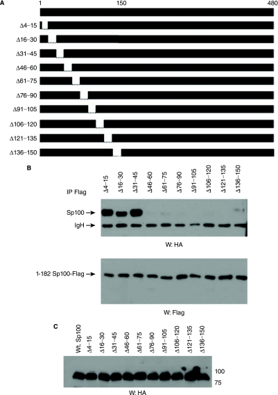
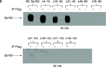
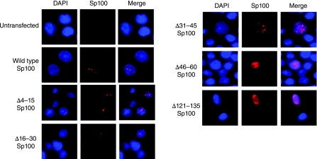
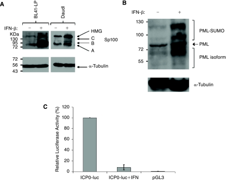
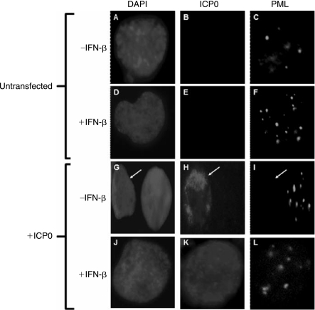
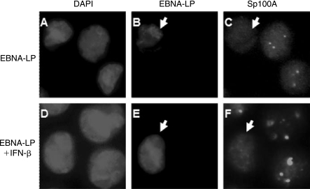
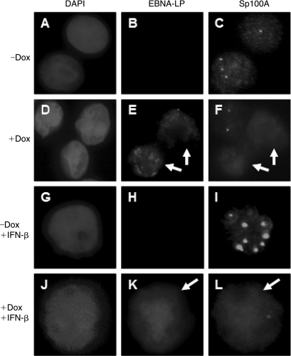
Epstein-Barr virus (EBV) efficiently immortalizes human B cells and is associated with several human malignancies. The EBV transcriptional activating protein EBNA2 and the EBNA2 coactivator EBNA-leader protein (EBNA-LP) are important for B cell immortalization. Recent observations from our laboratory indicate that EBNA-LP coactivation function is mediated through interactions with the interferon-inducible gene (ISG) Sp100, resulting in displacement from its normal location in promyelocytic leukemia nuclear bodies (PML NBs) into the nucleoplasm. The EBNA-LP- and interferon-mediated mechanisms that regulate Sp100 subnuclear localization and transcriptional function remain undefined. To clarify these issues, we generated a panel of Sp100 mutant proteins to ascertain whether EBNA-LP induces Sp100 displacement from PML NBs by interfering with Sp100 dimerization or through other domains. In addition, we tested EBNA-LP function in interferon-treated cells. Our results indicate that Sp100 dimerization, PML NB localization, and EBNA-LP interaction domains overlap significantly. We also show that IFN-beta does not inhibit EBNA-LP coactivation function. The results suggest that EBNA-LP might play a role in EBV-evasion of IFN-mediated antiviral responses.


Figures

FIG. 1.
FIG. 1.
Schematic of the Epstein-Barr Virus (EBV) genome and the sequence of EBNA-LP. The EBV genome is shown in linear form with relevant structural motifs and location of exons (black boxes) encoding latency proteins, which are labeled above. The internal repeat region (IR1) is also shown by the gray box. The viral transcript encoding EBNA-LP is shown below and is derived from the latency W promoter (Wp). The first exon transcribed from Wp is W0 and is noncoding. Alternative splicing from W0 to W1 can result in formation of an initiation codon (W1′) or no initiation codon (W1). Splicing that generates W1′ gives rise to transcripts encoding EBNA-LP. EBNA-LP is thus composed of repeated W1 and W2 exons as well as two unique exons known as Y1 and Y2. The transcript is bi-cistronic as it also contains the EBNA2 ORF at the 3′ end. The dashed line from the EBNA2 protein indicates promoters that are activated by EBNA2. The viral bidirectional promoter, which controls expression of LMP-1 and LMP2B, is indicated by asterisks. Transcripts encoding LMP-1 are shown below. A sequence comparison between EBV EBNA-LP and other nonhuman primate lymphocryptoviruses is shown below indicating presence of conserved regions (CR). Location of nuclear localization signals (NLS) and regions required for EBNA2 coactivation are indicated. Inverted triangles above the sequence indicate conserved serine residues.
FIG. 2.
FIG. 2.
Sp100A dimerization and EBNA-LP interaction domains are between amino acid residues 46–150. (A) Schematic representation of wild-type and mutant Sp100A. Boundaries of each deletion are indicated. (B) EBV-negative DG75 cells were cotransfected with 1–182 Sp100-Flag and wild-type or mutant Sp100A plasmids represented in (A). Following immunoprecipitation with anti-Flag antibodies, coprecipitated Sp100A was detected by Western blot using anti-HA antibodies. Anti-Flag antibodies were used to confirm 1–182-Flag precipitation. (C) Western blot detection of wild-type and mutant Sp100A proteins expressed in transfected DG75 cells. (D) DG75 cells were cotransfected with wild-type or mutant Sp100A and flag-tagged EBNA-LP expression plasmids. Immunoprecipitations and Western blots were carried out as outlined above, using anti-Flag and anti-HA antibodies.
FIG. 2.
FIG. 2.
Sp100A dimerization and EBNA-LP interaction domains are between amino acid residues 46–150. (A) Schematic representation of wild-type and mutant Sp100A. Boundaries of each deletion are indicated. (B) EBV-negative DG75 cells were cotransfected with 1–182 Sp100-Flag and wild-type or mutant Sp100A plasmids represented in (A). Following immunoprecipitation with anti-Flag antibodies, coprecipitated Sp100A was detected by Western blot using anti-HA antibodies. Anti-Flag antibodies were used to confirm 1–182-Flag precipitation. (C) Western blot detection of wild-type and mutant Sp100A proteins expressed in transfected DG75 cells. (D) DG75 cells were cotransfected with wild-type or mutant Sp100A and flag-tagged EBNA-LP expression plasmids. Immunoprecipitations and Western blots were carried out as outlined above, using anti-Flag and anti-HA antibodies.
FIG. 3.
FIG. 3.
The Sp100A PML NB targeting domain is between amino acid residues 46–150. Immunofluorescence detection of transiently expressed Sp100A proteins in DG75 cells. Sp100A (red) was monitored using mouse monoclonal anti-HA antibodies. Cells were co-stained with DAPI to detect the nucleus. Merged panels are shown in the far right hand column as indicated.
FIG. 4.
FIG. 4.
IFN-β regulates Sp100 and PML expression and the ICP0 promoter. (A) Western blot of endogenous Sp100A expression in BL41-LP and Daudi cells treated (+) or untreated (−) with 100 U/mL IFN-β for 24 h. Molecular weight markers are shown to the left side of the blot and Sp100 isoforms are indicated on the right side. α-Tubulin controls are shown in the panel below. (B) Western blot of endogenous PML expression in Daudi cells treated (+) or untreated (−) with 100 U/mL IFN-β for 24 h. Molecular weight markers are shown to the left side of the blot and PML isoforms indicated on the right side. α-Tubulin controls are shown in the panel below. (C) ICP0 promoter activity is inhibited by IFN-β. ICP0 promoter-driven luciferase plasmids were transfected into Daudi cells treated (+) and untreated (−) with IFN-β. A control sample was transfected with empty vector (pGL3). Relative luciferase activity for each of the transfections is indicated on the y-axis. Standard errors are indicated.
FIG. 5.
FIG. 5.
HSV-1 ICP0-mediated PML degradation is inhibited by IFN-β. Immunofluorescence detection of ICP0 and Sp100A in Daudi cells. Untransfected Daudi cells without IFN (−) or treated with IFN (+) for 24 h (+) are indicated on the left side of the panels. Daudi cells transfected with an ICP0 expression plasmid without IFN (−) or treated with IFN (+) for 24 h (+) are indicated similarly.
FIG. 6.
FIG. 6.
EBNA-LP displaces Sp100 from PML NBs in IFN-treated cells. Immunofluorescence of Daudi cells transfected with an EBNA-LP expression plasmid with or without IFN treatment. EBNA-LP and Sp100A were detected using monoclonal and polyclonal antibodies respectively as described in the Materials and Methods. DAPI staining of each field is shown in the panels on the left.
FIG. 7.
FIG. 7.
Induction of EBNA-LP expression in BL41-LP cells displaces Sp100A. BL41-LP cells were treated with 2 ng/mL doxycycline (Dox), resulting in the induction of EBNA-LP expression in about 50–80% of the cells. EBNA-LP and Sp100A were detected with monoclonal anti-EBNA-LP and polyclonal Sp100 antibodies respectively. Addition of Dox and/or IFN-β is shown on the left of each series of panels. White arrows indicate cells expressing EBNA-LP.
FIG. 8.
FIG. 8.
EBNA-LP coactivation function is not inhibited by IFN-β. Cp promoter luciferase reporter plasmids were transfected with EBNA2, EBNA-LP (LP), EBNA2 + LP, or EBNA2 + ΔCR3LP expression plasmids. Transfected cells were either untreated (A) or pulsed and maintained in 100 U/mL IFN-β for 24 h posttransfection (B). Relative fold-induction is shown on the left of each graph. Standard errors of the mean are indicated for each bar.

Similar articles

Cited by

References

    1. Adams A. Lidin B. Strander H. Cantell K. Spontaneous interferon-production and Epstein-Barr virus antigen expression in human lymphoid-cell lines. J Gen Virol. 1975a;28:219–223. - PubMed
    1. Adams A. Strander H. Cantell K. Sensitivity of Epstein-Barr Virus transformed human lymphoid-cell lines to interferon. J Gen Virol. 1975b;28:207–217. - PubMed
    1. Adamson AL. Kenney S. Epstein-Barr virus immediate-early protein BZLF1 is SUMO-1 modified and disrupts promyelocytic leukemia bodies. J Virol. 2001;75:2388–2399. - PMC - PubMed
    1. Ahn JH. Hayward GS. Disruption of PML-associated nuclear bodies by IE1 correlates with efficient early stages of viral gene expression and DNA replication in human cytomegalovirus infection. Virology. 2000;274:39–55. - PubMed
    1. Allday MJ. Crawford DH. Griffin BE. Epstein-Barr virus latent gene-expression during the initiation of B-cell immortalization. J Gen Virol. 1989;70:1755–1764. - PubMed

Publication types

MeSH terms