. 2008 Nov 15;314(19):3494-507.
doi: 10.1016/j.yexcr.2008.09.009. Epub 2008 Sep 25.

The mitochondrial outer membrane protein hFis1 regulates mitochondrial morphology and fission through self-interaction

Affiliations

The mitochondrial outer membrane protein hFis1 regulates mitochondrial morphology and fission through self-interaction

Madhavika N Serasinghe et al. Exp Cell Res. .

Erratum in

  • Exp Cell Res. 2009 Mar 10;315(5):899

Abstract

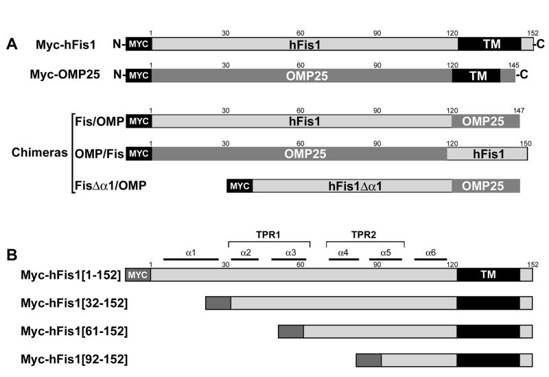
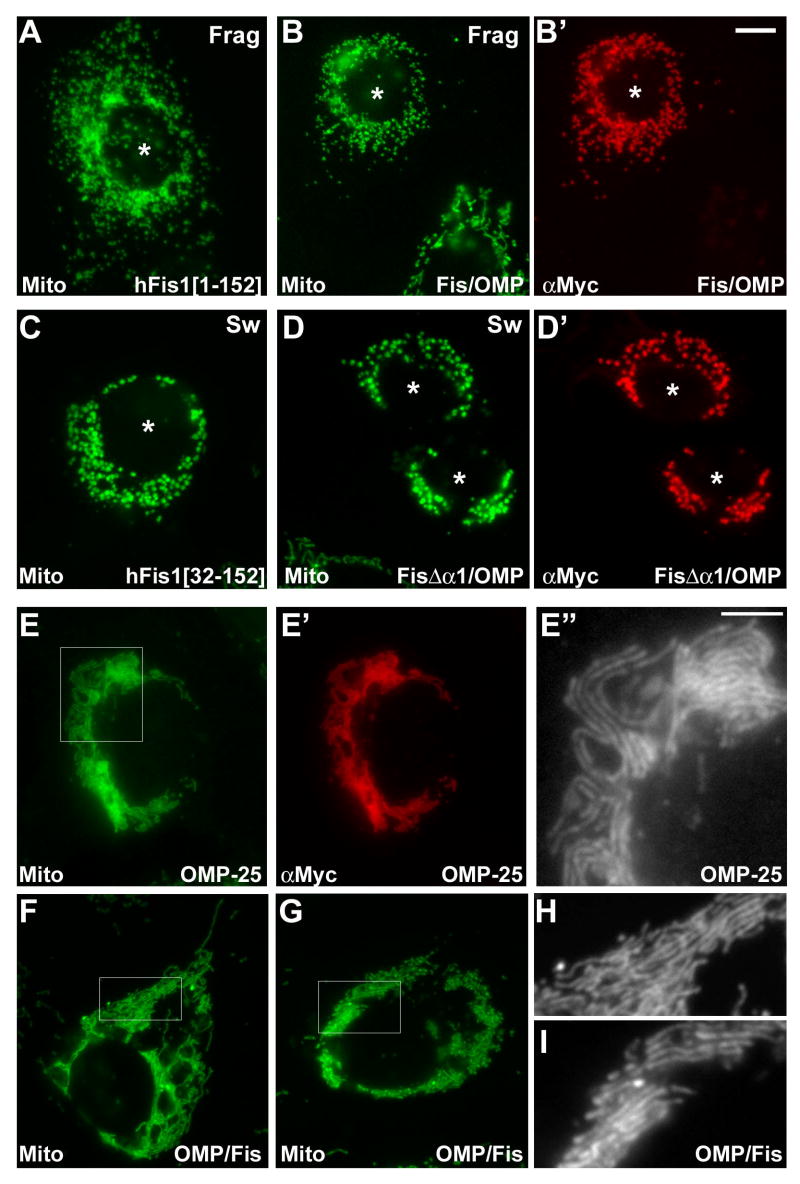
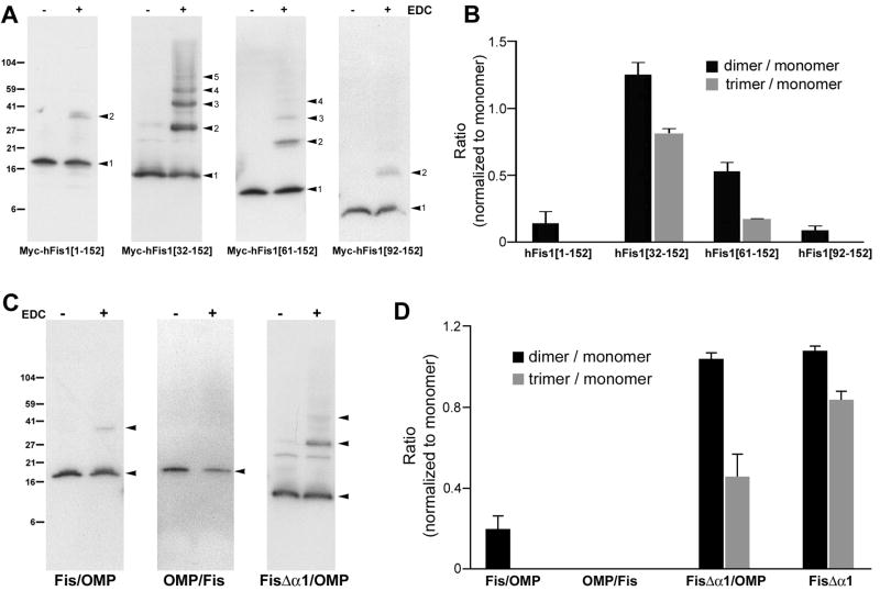
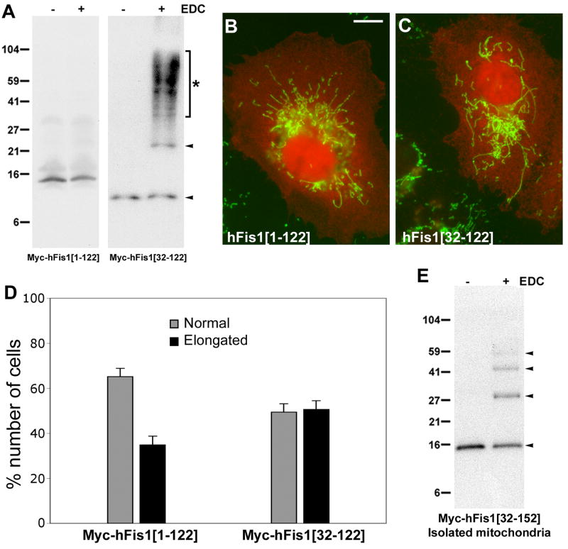
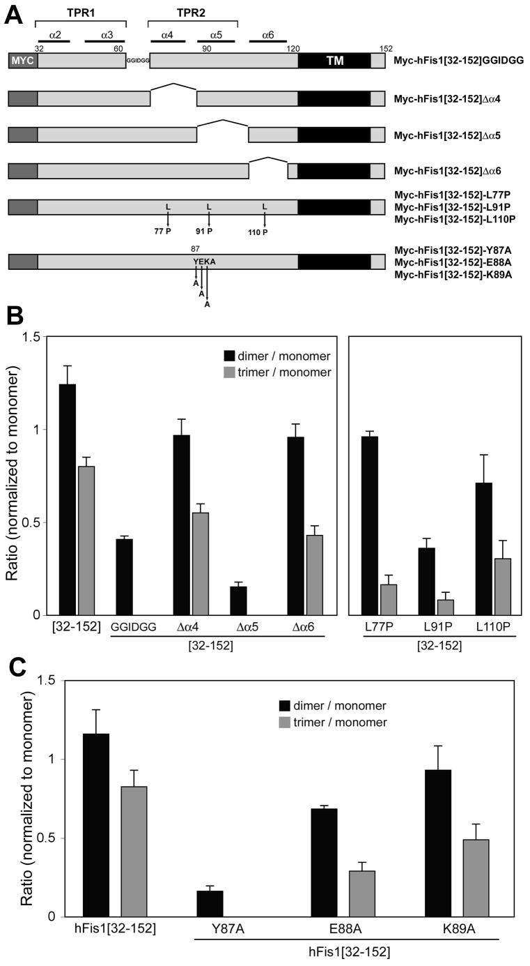
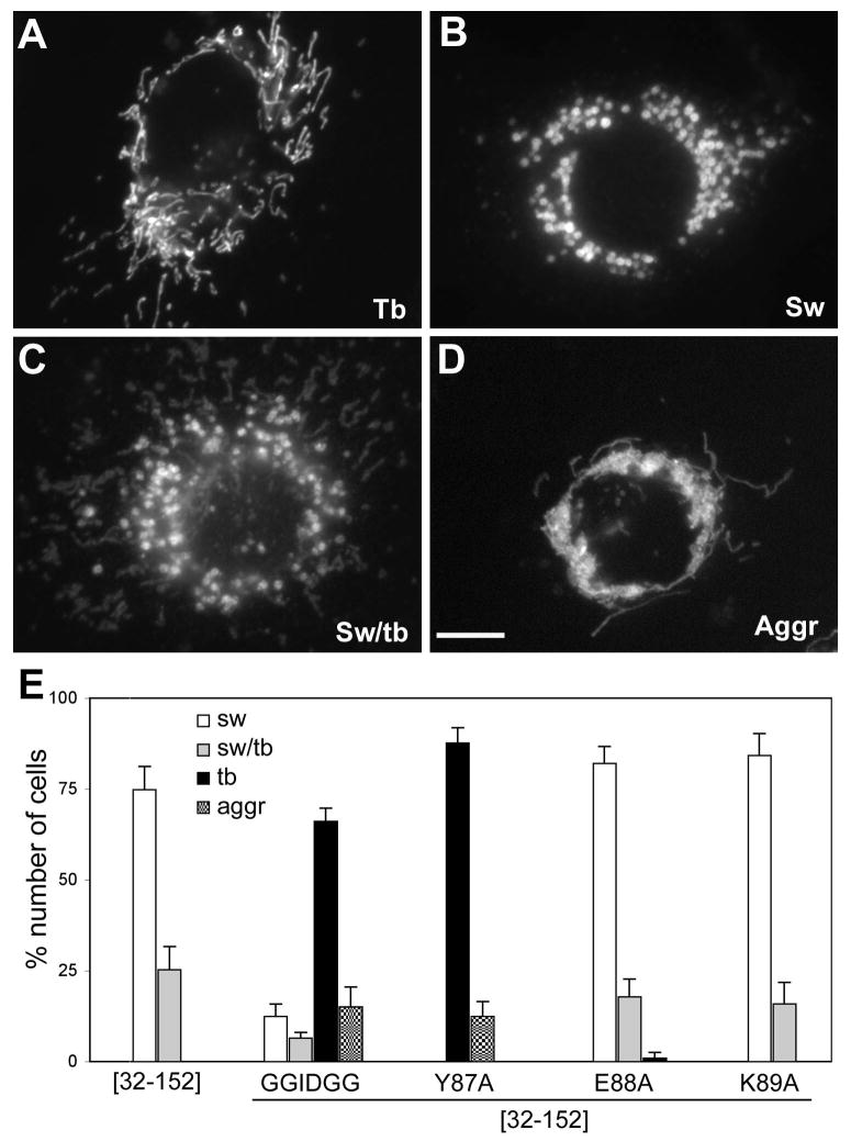
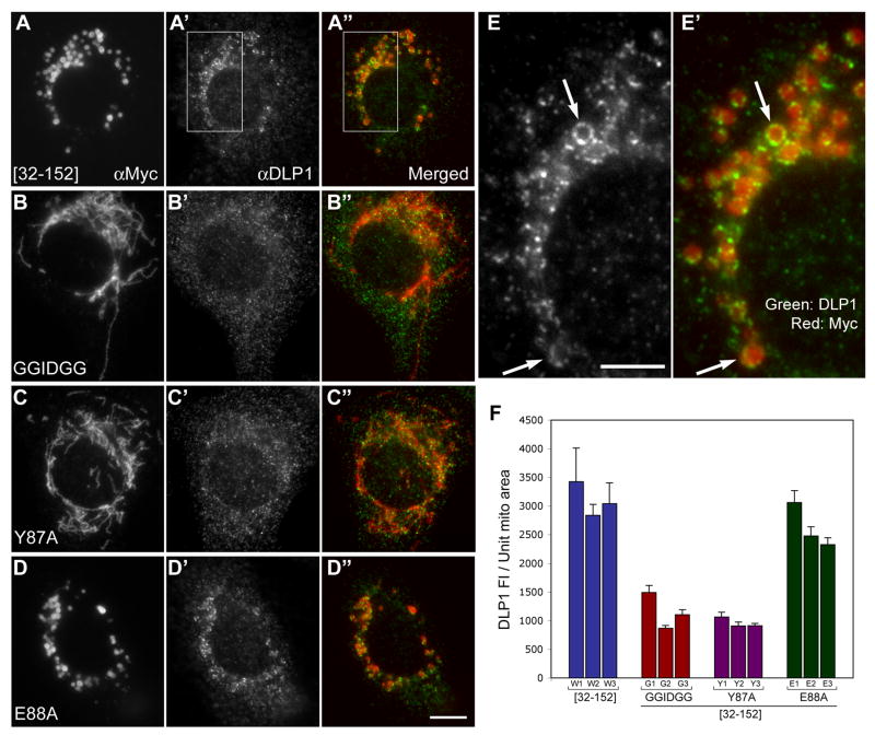
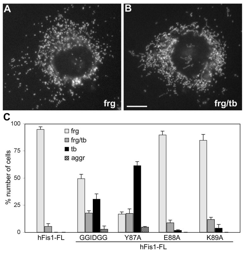
Mitochondrial fission in mammals is mediated by at least two proteins, DLP1/Drp1 and hFis1. DLP1 mediates the scission of mitochondrial membranes through GTP hydrolysis, and hFis1 is a putative DLP1 receptor anchored at the mitochondrial outer membrane by a C-terminal single transmembrane domain. The cytosolic domain of hFis1 contains six alpha-helices (alpha1-alpha6) out of which alpha2-alpha5 form two tetratricopeptide repeat (TPR) folds. In this study, by using chimeric constructs, we demonstrated that the cytosolic domain contains the necessary information for hFis1 function during mitochondrial fission. By using transient expression of different mutant forms of the hFis1 protein, we found that hFis1 self-interaction plays an important role in mitochondrial fission. Our results show that deletion of the alpha1 helix greatly increased the formation of dimeric and oligomeric forms of hFis1, indicating that alpha1 helix functions as a negative regulator of the hFis1 self-interaction. Further mutational approaches revealed that a tyrosine residue in the alpha5 helix and the linker between alpha3 and alpha4 helices participate in hFis1 oligomerization. Mutations causing oligomerization defect greatly reduced the ability to induce not only mitochondrial fragmentation by full-length hFis1 but also the formation of swollen ball-shaped mitochondria caused by alpha1-deleted hFis1. Our data suggest that oligomerization of hFis1 in the mitochondrial outer membrane plays a role in mitochondrial fission, potentially through participating in fission factor recruitment.


Figures

Figure 1
Figure 1. Chimeric and deletion constructs of hFis1 and OMP25
(A) Chimeric constructs between hFis1 and OMP25. The N-terminal cytosolic domain of hFis1 (aa 1-120) and the C-terminal transmembrane/tail region of OMP25 (aa 119-145) were fused to make the Fis/OMP. The converse chimera OMP/Fis was made of the cytosolic domain of OMP (aa 1-118) and the C-terminal region of hFis1 (aa 121-152). In FisΔα1/OMP, the α1 helix (aa 1-31) was deleted from the Fis/OMP chimera. (B) hFis1 full-length and N-terminal deletion constructs. Myc-hFis1[1-152] is the full length molecule. N-terminal 31, 60, and 91 amino acids were deleted in Myc-hFis1[32-152], Myc-hFis1[61-152], and Myc-hFis1[92-152], respectively. All constructs were tagged with the Myc-epitope at the N-terminus.
Figure 2
Figure 2. The N-terminal cytosolic domain of hFis1 contains the necessary information for hFis1-specific function
Chimeric proteins between hFis1 and OMP-25 were transfected into Clone 9 cells expressing GFP in mitochondria and immunostained with anti-Myc antibody. Fis/OMP that has the hFis1 cytosolic domain and the OMP-25 TM/C is localized to mitochondria (B′) and induces mitochondrial fragmentation (B), which is the same functional characteristics as the wild type hFis1 (A). (C) Overexpression of α1-deleted hFis1 (Myc-hFis1[32-152]) induces the formation of swollen ball-shape mitochondria. The chimera that has the α1 deletion in the Fis/OMP (FisΔα1/OMP) causes the same swollen mitochondrial morphology (D′) and correctly distributes to the mitochondria (D). Asterisks in A-D′ denote transfected cells. Myc-OMP-25 is localized to mitochondria (E′) and induces perinuclear clustering of mitochondria (E). Higher magnification image show that mitochondrial tubules are aligned and stacked (E″). (F, G, H, I) The chimera that has the OMP-25 cytosolic domain and hFis1 TM/C induces the same mitochondrial phenotype as the wild type Myc-OMP-25. Panels E″, H, and I are enlarged images of the boxed regions in E, F, and G, respectively. The scale bar in the panel E″ (5 μm) represents the magnification of the images E″, H, and I, whereas the one in B′ (10 μm) is for the rest of the images.
Figure 3
Figure 3. hFis1 forms oligomers in an α1-helix dependent manner and the oligomerization occurs within the cytosolic domain
(A) EDC crosslinking of cells transiently expressing different Myc-tagged hFis1 constructs. Cell lysates were separated by SDS-gel electrophoresis and hFis1 proteins were detected by anti-Myc immunoblotting. Myc-hFis1[32-152] and Myc-hFis1[61-152] show oligomeric complexes whereas Myc-hFis1[1-152] and Myc-hFis1[92-152] showed only a small amount of dimer. (B) Densitometric analyses of the hFis1 oligomerization. Monomeric, dimeric, and trimeric bands were subjected to densitometry, and densities of dimer and trimer were normalized to that of the monomeric band. Results from three different experiments were analyzed. Error bars represent SEM. (C) EDC crosslinking of cells expressing the chimeric constructs. OMP/Fis shows no complex formation whereas a small amount of dimer is seen in Fis/OMP. The α1-deleted chimera FisΔα1/OMP shows an increased dimer and trimer formation. (D) Densitometric analyses for oligomerization of chimeras. The same method described in (B) was used.
Figure 4
Figure 4. Membrane anchoring of hFis1 is necessary for oligomerization
(A) Crosslinking of Myc-hFis1[1-122] and Myc-hFis1[32-122]. The lack of the C-terminal transmembrane and tail domain in these constructs results in the loss of membrane anchoring. No crosslinked bands were detected with Myc-hFis1[1-122] whereas a high molecular weight smear between the 27 and 104 kD markers (asterisk) was seen upon crosslinking of cells expressing Myc-hFis1[32-122]. (B, C) Clone 9 cells harboring GFP in mitochondria were transfected with Myc-hFis1[1-122] (B) and Myc-hFis1[32-122] (C), and immunofluorescence was performed using anti-Myc antibodies. Both mutants showed diffuse cytosolic distribution (red fluorescence). Mitochondrial morphologies were tubular with both mutants, indicating the loss of the ability to induce fragmentation and swelling. Mitochondria in many transfected cells were elongated and entangled (C), indicating blocked fission. Scale bar: 10 μm. (D) Cell counting for mitochondrial morphology shows that 35 and 50% of cells transfected with Myc-hFis1[1-122] and Myc-hFis1[32-122], respectively, contain elongated mitochondria. (E) Crosslinking of the mitochondrial fraction isolated from cells expressing Myc-hFis1[32-152]. Mitochondria were isolated by differential centrifugation. Isolated mitochondria were resuspended in the crosslinking buffer and subjected to the EDC crosslinking. Dimeric, trimeric, and tetrameric bands were detected, indicating the presence of oligomeric forms of hFis1.
Figure 5
Figure 5. Identification of hFis1 regions participating in oligomerization
(A) Internal deletion and point mutants used for crosslinking experiments. In Myc-hFis1[32-152]GGIDGG, the loop sequence KGSKEEQ that links the α3 and α4 helices was substituted with GGIDGG. Amino acids [71-86], [87-104], and [105-118] were deleted in Myc-hFis1[32-152]Δα4, Myc-hFis1[32-152]Δα5, Myc-hFis1[32-152]Δα6, respectively. Helix-breaking L-to-P mutations (L77P, L91P, and L110P) were made in α4, α5, and α6 helices. The three amino acids in the α5-helix were mutated to alanine (Y87A, E88A, and K89A). (B, C) Densitometric analyses of hFis1 oligomeric bands for crosslinking experiments. Dimeric and trimeric band densities were normalized against the monomer density. Results from three different experiments were analyzed. Error bars represent SEM. Myc-hFis1[32-152]GGIDGG and Myc-hFis1[32-152]Δα5 showed a marked decrease in oligomerization whereas Δα4 and Δα6 formed dimers and trimers to the extent comparable to Myc-hFis1[32-152] (B). The α5 helix-breaking mutation (L91P) also decreased oligomerization, but the ones in the α4 and α6 did not (B). No trimer formation and very little dimer were observed with Myc-hFis1[32-152]-Y87A (C), indicating that the tyrosine residue in the α5 helix plays a role in hFis1 oligomerization.
Figure 6
Figure 6. Mitochondrial swelling is abolished in cells expressing oligomerization-defective mutants
Clone 9 cells harboring GFP in the mitochondrial matrix were transfected with different constructs, and mitochondrial morphologies were evaluated. (A, B, C, D) Different mitochondrial morphologies: normal tubular morphology (A, Tb), swollen ball-shape mitochondria (B, Sw), The mixture of swollen and tubular mitochondria (C, Sw/tb), and aggregated and collapsed mitochondria (D, Aggr). Scale bar: 10μm. (E) Cell counting for different mitochondrial phenotypes. More than 300 transfected cells were examined from three separate experiments. Most of the cells overexpressing Myc-hFis1[32-152] contained swollen mitochondria or a mixture of swollen and tubular mitochondria. These phenotypes were mostly absent in cells transfected with the oligomerization defective mutants Myc-hFis1[32-152]GGIDGG and Myc-hFis1[32-152]-Y87A, but not with E88A and K89A mutants. The normal tubular mitochondrial morphology was predominant in cells transfected with the oligomerization-defective mutants. Some cells showed the aggregated and entangled mitochondrial phenotype. Error bars represent SEM.
Figure 7
Figure 7. Oligomerization-defective mutations abolish the ability of the α1-deleted hFis1 to increase the DLP1 association to the mitochondria
Indirect immunofluorescence was carried out for DLP1 and the Myc epitope in cells transfected with Myc-hFis1[32-152] (A, A', A″, E. E′), Myc-hFis1[32-152]-GGIDGG (B, B', B″), Myc-hFis1[32-152]-Y87A (C, C', C″), and Myc-hFis1[32-152]-E88A (D, D', D″). (A, A', A″) Myc-hFis1[32-152] is localized to the swollen mitochondria (A) brighter DLP1 puncta are concentrated on and around the ball-shape mitochondria (A′). DLP1 forms patches and often coats the entire circumference of swollen mitochondria (arrows in E, E′). (B, B', B″, C, C', C″) No significant DLP1 association is apparent with mitochondrial tubules stained for Myc-hFis1[32-152]-GGIDGG (B, B', B″) and Myc-hFis1[32-152]-Y87A (C, C', C″). (D, D', D″) Myc-hFis1[32-152]-E88A mutant show the DLP1 staining pattern around the swollen mitochondria (D', D″) similar to the Myc-hFis1[32-152]. (E, E′) Enlarged images of boxed regions in A′ and A″, respectively. The scale bar in the panel E (5 μm) represents the magnification of the images E and E′ whereas the one in D″ (10 μm) is for the rest of the images. (F) Quantification of DLP1 association with mitochondria. 5 to 11 isolated, discernable mitochondria were selected from each cell and the mitochondrial area was measured. Total DLP1 fluorescence associated with a given mitochondrion was measured and normalized against the area of the same mitochondrion to calculate the amount of mitochondria-associated DLP1 per unit mitochondrial area. The graph shows the measurements of three individual cells from Myc-hFis1[32-152] (W1-W3), Myc-hFis1[32-152]-GGIDGG (G1-G3), Myc-hFis1[32-152]-Y87A (Y1-Y3), and Myc-hFis1[32-152]-E88A (E1-E3). An approximately three-fold reduction of mitochondria-associated DLP1 fluorescence was observed in oligomerization-defective mutants compared to Myc-hFis1[32-152] and Myc-hFis1[32-152]-E88A. Error bars represent SEM.
Figure 8
Figure 8. Oligomerization-defective mutants have a diminished ability to induce mitochondrial fragmentation
Mitochondrial morphologies were examined in cells transfected with different hFis1 constructs. Overexpression of full-length Myc-tagged wild type hFis1 caused the fragmented mitochondrial phenotype (A, frag). Some cells contained a mixture of fragmented and tubular mitochondria (B, frg/tb). Scale bar: 10μm. (C) Cell counting of more than 300 cells transfected with each construct was performed in three experiments. Decreased numbers of cells showed fragmented mitochondria among cells transfected with the oligomerization-defective mutants, GGIDGG and Y87A. Myc-hFis1-E88A and K89A had no significant effect on mitochondrial fragmentation upon overexpression. Error bars represent SEM.

Similar articles

Cited by

References

    1. Yoon Y. Regulation of mitochondrial dynamics: Another process modulated by Ca2+ signal? Sci STKE. 2005;2005:pe18. - PubMed
    1. Yoon Y. Sharpening the scissors: mitochondrial fission with aid. Cell Biochem Biophys. 2004;41:193–205. - PubMed
    1. Bereiter-Hahn J. Behavior of mitochondria in the living cell. Int Rev Cytol. 1990;122:1–63. - PubMed
    1. Sesaki H, Jensen RE. Division versus fusion: Dnm1p and Fzo1p antagonistically regulate mitochondrial shape. J Cell Biol. 1999;147:699–706. - PMC - PubMed
    1. Alexander C, Votruba M, Pesch UE, Thiselton DL, Mayer S, Moore A, Rodriguez M, Kellner U, Leo-Kottler B, Auburger G, Bhattacharya SS, Wissinger B. OPA1, encoding a dynamin-related GTPase, is mutated in autosomal dominant optic atrophy linked to chromosome 3q28. Nat Genet. 2000;26:211–215. - PubMed

Publication types

MeSH terms