. 2009 Jan;130(1):98-109.
doi: 10.1016/j.clim.2008.08.025. Epub 2008 Oct 8.

Can oligomeric T-cell receptor be used as a tool to detect viral peptide epitopes on infected cells?

Affiliations

Can oligomeric T-cell receptor be used as a tool to detect viral peptide epitopes on infected cells?

Nadia Anikeeva et al. Clin Immunol. 2009 Jan.

Abstract

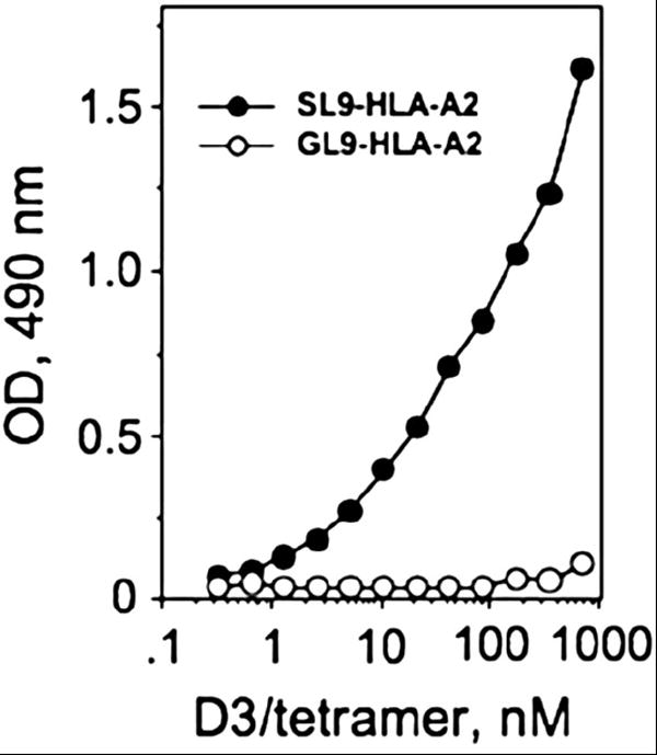
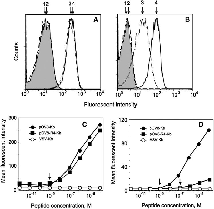
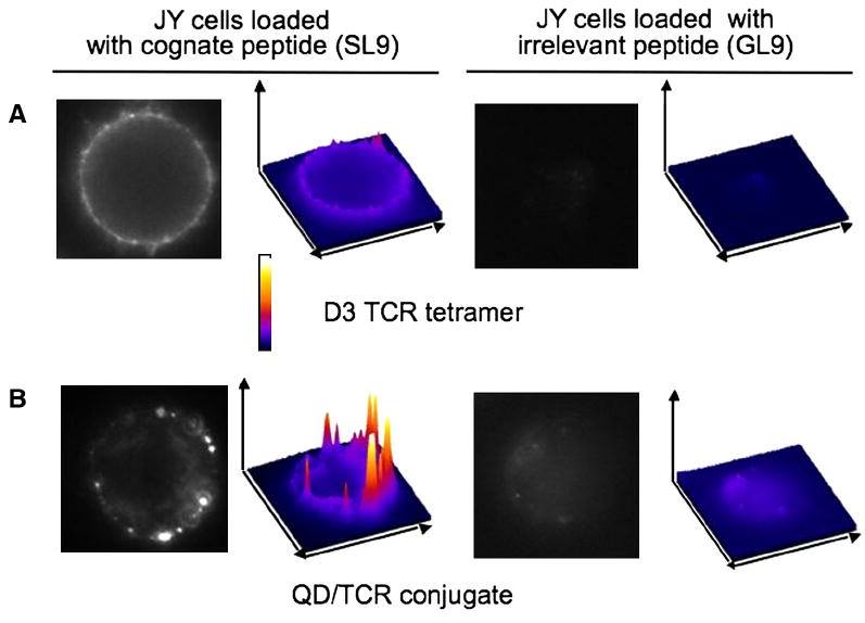
We have utilized soluble HIV Gag-specific T-cell receptor (TCR) D3 with low affinity and TCR-like antibody 25-D1.16 recognizing its natural peptide-MHC (pMHC) ligand with high affinity to determine how affinity and off-rate of the receptor-pMHC interactions affect the sensitivity of pMHC detection on the cell surface. We found that with soluble TCR cognate pMHCs can be detected only at relatively high cell surface densities when the TCR was oligomerized using either Streptavidin or quantum dot (QD) scaffolds. While the higher affinity probe led to a greater sensitivity of pMHC detection, monomers and oligomers of the probe showed essentially the same detection limit, which is restricted by the sensitivity of standard flow cytometry technique. We have also shown that imaging of QD/TCR specifically bound to cognate pMHC on the cell surface yielded a very bright fluorescent signal that can enhance the sensitivity of viral peptide detection on infected cells.


Figures

Figure 1
Figure 1. Assembly of D3/TCR tetramer
SDS PAGE at non-reducing condition shows that all biotinylated D3 were bound to Streptavidin after D3/tetramer assembly. Although trimer (Strepavidin + 3 molecules of D3) dominates the tetramer preparation, other species (monomer, dimer, and tetramer) were present as well (Lane 1); small amount of aggregates was also evident (upper band in Lane 1). Lane 2: Streptavidin; lane 3: D3 after freezing and storage at -70°C (aggregates and a small amount of degradation products are visible); lane 4: freshly isolated D3 TCR.
Fig. 2
Fig. 2. D3/tetramer specifically bound to immobilized SL9-HLA-A2 but not GL9-HLA-A2 complex
Flat-bottom 96-well plate was covered overnight (4°C) by soluble peptide-HLA-A2 complex and the plates were blocked with 1% BSA/PBS. D3/tetramer was then added at various concentrations and the amount of bound tetramer was quantified by measuring OD at 490 nm. Indicated concentrations refer to the concentration of D3 in the preparation of the tetramer.
Fig. 3
Fig. 3. D3/tetramer specifically stains HLA-A2+ JY cells sensitized with HIV gag-derived peptide SLYNTVATL (SL9) but not with GILGFVFTL (GL9) peptide from influenza virus
The cells (106/ml in R10) were incubated with respected peptides at 100 μM 60 minutes at 37°C. To some samples soluble β2m was added at 100 μg/ml. The culture supernatant was removed by centrifugation, and the cells were stained with 5 μM D3 TCR tetramer: (1), intact cells; (2), cells sensitized with irrelevant GL9 peptide; (3) cells sensitized with cognate SL9 peptide; (4) cells sensitized with cognate SL9 peptide in the presence of β2m. The presence of soluble β2m facilitated staining of target cells bearing cognate pMHC complexes by soluble fluorescence-labeled TCR/tetramer. Staining of the cells sensitized with Flu-derived peptide GL9 was indistinguishable from the negative control.
Fig. 4
Fig. 4. Enzymatic amplification of fluorescent signal results in profound increase of the staining intensity, while the sensitivity of the epitope detection was similar to the standard staining procedure
A. Intact JY cells (1) and JY cells bearing either cognate SL9-HLA-A2 (3) or irrelevant GL9-HLA-A2 (2) complexes stained with 0.2 μM of PE-labeled D3/tetramer. B. The same cells stained with HRP-labeled D3/tetramer at TCR concentration 0.2 μM followed by tyramine-based signal amplification technique (see Material and Methods). C. JY cells were sensitized with indicated concentration of the SL9 or GL9 peptides and were stained with D3/tetramer using the amplification technique.
Fig. 5
Fig. 5. Multivalent QD/D3 TCR conjugate shows similar specificity and sensitivity as the D3 TCR/tetramer
QD/TCR conjugates specifically stain HLA-A2+ JY cells sensitized with cognate SL9 (3). Staining of cells sensitized with control GL9 peptide (2) was compared with unlabeled cells (1). A. JY cells bearing with SL9-HLA-A2 (3) or GL9-HLA-A2 (2) complexes stained with QD(520)/TCR conjugate (0.5 μM of QDs) accommodating 10 TCR molecules per dot. B. The same cells stained with QD(620)/TCR conjugate (20 nM of QDs) accommodating 100 TCR molecules per dot. C. JY cells were sensitized with indicated concentration of the SL9 or GL9 peptide and were stained with QD(620)/TCR conjugate at QDs concentration 20 nM.
Fig. 6
Fig. 6. The sensitivity of pOV8-Kb detection with monomeric and tetrameric 25-D1.16 Fab are very similar, while the staining for pOV8-R4-Kb with the tetramer is more sensitive than with monomeric reagent
25-D1.16 Fab tetramer (A) and 25-D1.16 Fab monomer (B) specifically stain EL-4 cells pulsed with 10-4 M pOV8 (4, solid line) and pOV8-R4 (3, dotted line). Staining of EL4 cells with irrelevant peptide VSV (2, dashed line) was not different from self fluorescence of intact cells (1, shown in grey). The intensity of the staining with the tetramer was somewhat higher than with the monomeric Fab; slight difference in the background staining was evident. EL-4 cells were pulsed with either strong agonist pOV8 or weak agonist pOV8-R4 or a null peptide VSV at various concentrations and were stained with 25-D1.16 Fab tetramer (C) or 25-D1.16 Fab monomer (D) at 40 nM concentration of the Fab.
Fig. 7
Fig. 7. QD/TCR conjugate is a better reagent for pMHC epitope detection by fluorescent microscopy as compared to D3 TCR/tetramer
HLA-A2+ JY cells sensitized with SL9-HLA-A2 at peptide concentration 10-4 M were stained with D3/tetramer/PE (A) and QD/D3 TCR (B). Staining of the cells sensitized with control peptide GL9 was not evident. The staining pattern with D3 tetramer was less intense and more diffuse as compared to the QD/D3 TCR staining. Surface plots of the staining were built with ImageJ software.

Similar articles

Cited by

References

    1. Eisen HN, Sykulev Y, Tsomides TJ. The antigen-specific T-cell receptor and its reactions with peptide-MHC complexes. In: Haber E, editor. Antigen-Binding Molecules Antibodies and T-cell Receptors. Academic Press; San Diego: 1997. pp. 1–56.
    1. Davis MM, Boniface JJ, Reich Z, Lyons D, Hampl J, Arden B, Chien Y. Ligand recognition by alpha beta T cell receptors. Annual Review of Immunology. 1998;16:523–44. - PubMed
    1. Sykulev Y, Anikeeva N, Kalams SA. Soluble TCR/tetramer as a tool to study the distribution of immunodominant HIV Gag peptide epitope on the surface of target cells. Experimental Biology Meeting; New Orlean, LA. 2002.
    1. Subbramanian RA, Moriya C, Martin KL, Peyerl FW, Hasegawa A, Naoi A, Chhay H, Autissier P, Gorgone DA, Lifton MA, Kuus-Reichel K, Schmitz JE, Letvin NL, Kuroda MJ. Engineered T-cell receptor tetramers bind MHC-peptide complexes with high affinity. Nat Biotechnol. 2004;22:1429–34. - PubMed
    1. Laugel B, Boulter JM, Lissin N, Vuidepot A, Li Y, Gostick E, Crotty LE, Douek DC, Hemelaar J, Price DA, Jakobsen BK, Sewell AK. Design of soluble recombinant T cell receptors for antigen targeting and T cell inhibition. J Biol Chem. 2005;280:1882–92. - PubMed

Publication types

MeSH terms