. 2009 Feb;14(1):2-7.
doi: 10.1016/j.siny.2008.08.011. Epub 2008 Oct 8.

Prenatal inflammation and lung development

Affiliations

Prenatal inflammation and lung development

Boris W Kramer et al. Semin Fetal Neonatal Med. 2009 Feb.

Abstract

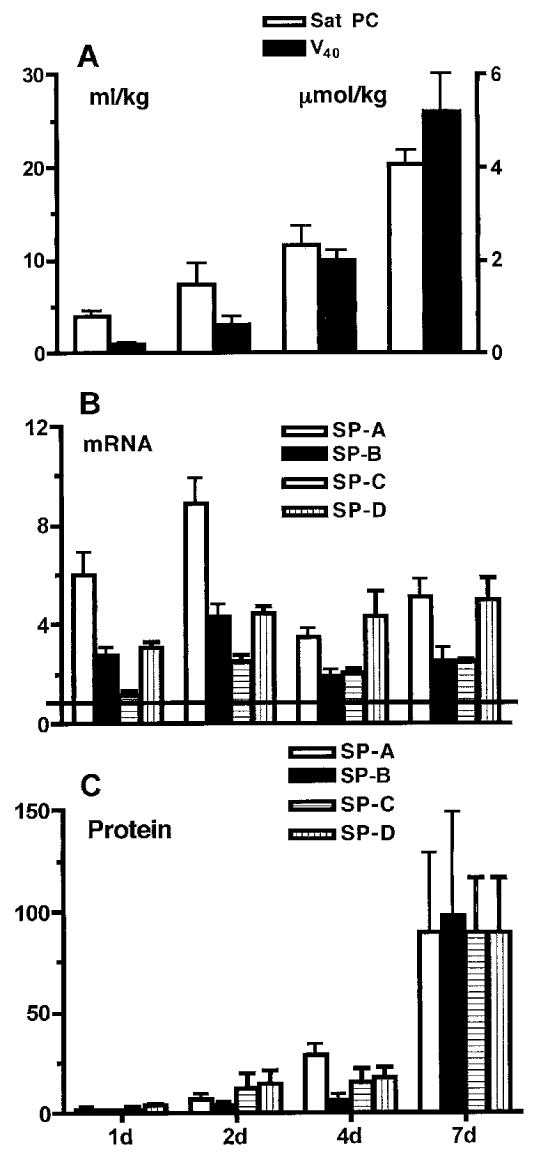
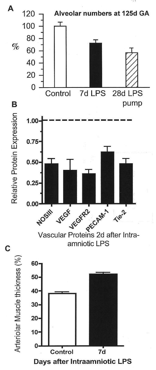
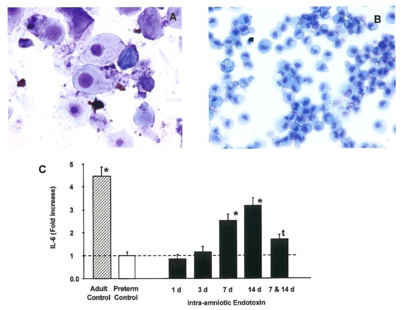
Prenatal exposure of very low birth weight infants to chronic indolent chorioamnionitis with organisms such as mycoplasma and ureaplasma is frequent. Chorioamnionitis is inconsistently associated with changed risks of respiratory distress syndrome (RDS) or bronchopulmonary dysplasia (BPD), probably because the diagnosis of chorioamnionitis does not quantify the extent or duration of the fetal exposures to infection and inflammation. The correlations between prenatal exposures and postnatal lung disease also are confounded by the imprecision of the diagnoses of RDS and BPD. In animal models, chorioamnionitis caused by pro-inflammatory mediators or live ureaplasma induces lung maturation, but also causes alveolar simplification and vascular injury. Intra-amniotic endotoxin administration also modulates the fetal innate immune system, resulting in maturation of monocytes to alveolar macrophages and the induction or paralysis of inflammatory responses depending on exposure history. Prenatal inflammation can have profound effects on the fetal lung and subsequent immune responses.


Conflict of interest statement

Conflict of interest statement: None declared.

Figures

Figure 1
Figure 1
Surfactant proteins (SP) and lipids increase as lung function improves following intra-amniotic endotoxin. Fetal sheep were exposed to 20 mg E. coli endotoxin given by intra-amniotic injection at the intervals given on the horizontal axis prior to preterm delivery at 125 d gestation. (A) Lung gas volume (V40; solid bars) measured as mL/kg air at 40 cm pressure increased over 7 d, as did the amount of saturated phosphatidylcholine (Sat PC, μmol/kg; hollow bars). (B) The amount of mRNA for the surfactant proteins in lung tissue also increased and remained elevated relative to the normalised values of 1 for lung tissue from lambs not exposed to endotoxin. (C) The amounts of the surfactant proteins in bronchoalveolar lavage increased from the normalised value of 1 for lambs not exposed to intra-amniotic endotoxin. Data calculated and redrawn from Ref. 24. (B) and (C): hollow bars, SP-A; solid bars, SP-B; hatched bars, SP-C; stippled bars, SP-D.
Figure 2
Figure 2
Intra-amniotic endotoxin inhibits alveolarisation and causes vascular injury. Preterm fetal sheep were given intra-amniotic endotoxin (lipopolysaccharide, LPS). (A) Compared with controls, alveolar numbers decreased 7 d after intra-amniotic LPS or 28 d after exposure to a continuous intra-amniotic LPS infusion. (B) Expression of several vascular proteins by western blot decreased 2 d after intra-amniotic LPS relative to controls (dashed line). (C) Distal arteriolar smooth muscle thickness increased 7 d after intra-amniotic LPS compared with controls. Data redrawn from Refs 25, and . GA, gestational age; NOS, nitric oxide synthase; VEGF, vascular endothelial growth factor; VEGFR, VEGF receptor; PECAM, platelet endothelial cell adhesion molecule.
Figure 3
Figure 3
Appearance of alveolar macrophages and interleukin (IL)-6 production by lung monocytes. (A) The bronchoalveolar lavage of a control fetal lamb contains only epithelial cells and occasional monocytes. (B) Seven days after intra-amniotic endotoxin the cells are mature-appearing monocytes and neutrophils. (C) Monocytic cells were recovered from fetal lungs at periods from 1 to 14 d after intra-amniotic endotoxin and challenged in culture with endotoxin. Alveolar macrophages from adult sheep were the adult control cells. The fetal lung monocytes produced very little IL-6 relative to the alveolar macrophages from the adult. The fetal cells produced IL-6 in response to in-vitro challenge 7 and 14 d after intra-amniotic endotoxin. However, exposure to intra-amniotic endotoxin 7 and 14 d before in-vitro endotoxin challenge resulted in decreased responsiveness. Data from experiments reported by Kramer et al., *P < 0.05 vs preterm control; tP < 0.05 vs 14 d endotoxin.

References

    1. Charafeddine L, D'Angio CT, Phelps DL. Atypical chronic lung disease patterns in neonates. Pediatrics. 1999;103:759–65. - PubMed
    1. Preterm birth – causes, consequences and prevention. Institute of Medicine. Washington: National Academic Press; 2007.
    1. Romero R, Gomez R, Ghezzi F, et al. A fetal systemic inflammatory response is followed by the spontaneous onset of preterm parturition. Am J Obstet Gynecol. 1998;179:186–93. - PubMed
    1. Perni SC, Vardhana S, Korneeva I, et al. Mycoplasma hominis and Ureaplasma urealyticum in midtrimester amniotic fluid: association with amniotic fluid cytokine levels and pregnancy outcome. Am J Obstet Gynecol. 2004;191:1382–6. - PubMed
    1. Gerber S, Vial Y, Hohlfeld P, Witkin SS. Detection of Ureaplasma urealyticum in second-trimester amniotic fluid by polymerase chain reaction correlates with subsequent preterm labor and delivery. J Infect Dis. 2003;187:518–21. - PubMed

Publication types