. 2008 Dec 12;283(50):34667-76.
doi: 10.1074/jbc.M805624200. Epub 2008 Oct 9.

Elevated synaptic activity preconditions neurons against an in vitro model of ischemia

Affiliations

Elevated synaptic activity preconditions neurons against an in vitro model of ischemia

Joseph S Tauskela et al. J Biol Chem. .

Abstract

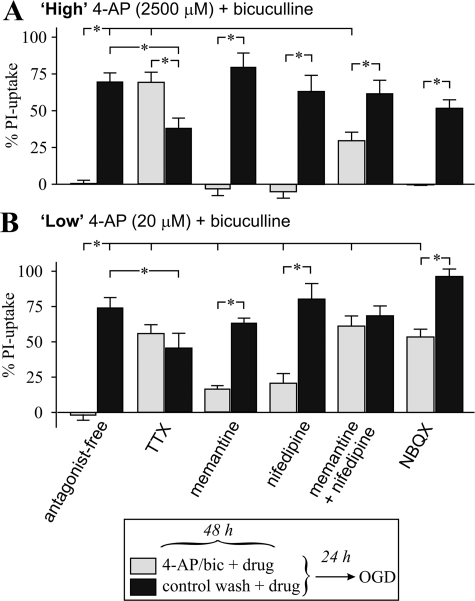
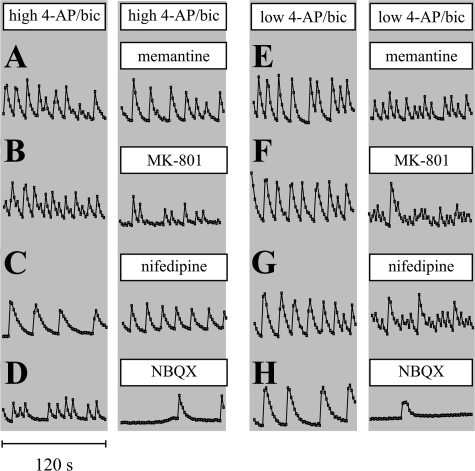
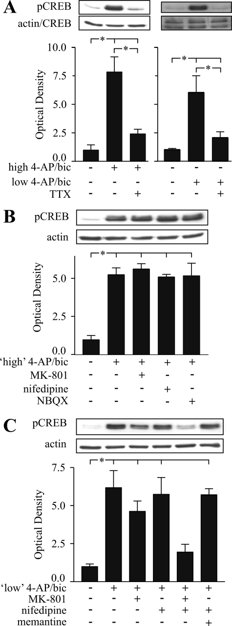
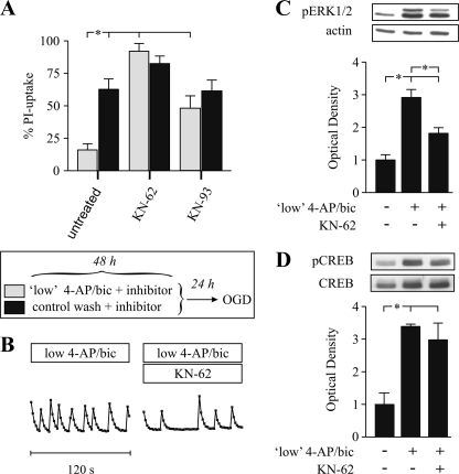
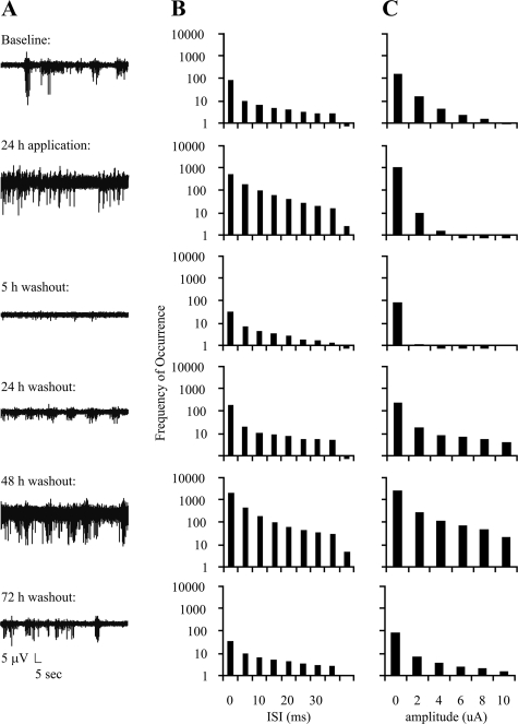
Tolerance to otherwise lethal cerebral ischemia in vivo or to oxygen-glucose deprivation (OGD) in vitro can be induced by prior transient exposure to N-methyl-D-aspartic acid (NMDA): preconditioning in this manner activates extrasynaptic and synaptic NMDA receptors and can require bringing neurons to the "brink of death." We considered if this stressful requirement could be minimized by the stimulation of primarily synaptic NMDA receptors. Subjecting cultured cortical neurons to prolonged elevations in electrical activity induced tolerance to OGD. Specifically, exposing cultures to a K(+)-channel blocker, 4-aminopyridine (20-2500 microm), and a GABA(A) receptor antagonist, bicuculline (50 microm) (4-AP/bic), for 1-2 days resulted in potent tolerance to normally lethal OGD applied up to 3 days later. Preconditioning induced phosphorylation of ERK1/2 and CREB which, along with Ca(2+) spiking and OGD tolerance, was eliminated by tetrodotoxin. Antagonists of NMDA receptors or L-type voltage-gated Ca(2+) channels (L-VGCCs) applied during preconditioning decreased Ca(2+) spiking, phosphorylation of ERK1/2 and CREB, and OGD tolerance more effectively when combined, particularly at the lowest 4-AP concentration. Inhibiting ERK1/2 or Ca(2+)/calmodulin-dependent protein kinases (CaMKs) also reduced Ca(2+) spiking and OGD tolerance. Preconditioning resulted in altered neuronal excitability for up to 3 days following 4-AP/bic washout, based on field potential recordings obtained from neurons cultured on 64-channel multielectrode arrays. Taken together, the data are consistent with action potential-driven co-activation of primarily synaptic NMDA receptors and L-VGCCs, resulting in parallel phosphorylation of ERK1/2 and CREB and involvement of CaMKs, culminating in a potent, prolonged but reversible, OGD-tolerant phenotype.


Figures

FIGURE 1.
FIGURE 1.
A–H, preconditioning rat cortical neurons with 4-aminopyridine/bicuculline induces tolerance to otherwise lethal OGD. Neurons were subjected to sham wash or to preconditioning by 2.5 mm 4-AP plus 50 μm bicuculline (4-AP/bic) for 48 h, followed by a 24-h recovery period, then subjected to control wash or 65–80 min OGD, followed by assessment of neurotoxicity by measurement of the % PI-uptake 24 h later. Same-field phase contrast and PI fluorescence image pairs were acquired from the following conditions: A and B, sham wash; C and D, 4-P/bic preconditioning, followed by OGD; E and F, OGD; or G and H, 100% neuronal kill achieved by subjecting sister cultures to 100 μm NMDA for 15 min. I, preconditioning increases neuronal activity. Neuronal intracellular Ca2+ spiking in Fluo-4-loaded neurons 0.5–2.5 h following application of sham wash, 50 μm bicuculline (bic), 20 μm 4-aminopyridine plus 50 μm bicuculline (4-AP/bic), or to 4AP/bic applied with a Na+ channel blocker, tetrodotoxin (TTX, 1 μm). J–P, a wide range of 4-AP/bic concentrations and duration of exposure, as well as the duration of the recovery interval between preconditioning and OGD, result in neuroprotection. Cultures were subjected to sham wash or to preconditioning with 50 μm bicuculline combined with 4-AP (20, 300, or 2500 μm) for 4–48 h, followed by washout and recovery for 0–96 h, subsequently exposed to 65–80 min OGD, followed by measurement of % PI-uptake 24 h later (n = 12–18). Preconditioning resulted in a significant suppression of % PI-uptake following OGD (p < 0.05; calculated using an ANOVA), except for the shortest 4-AP/bic duration investigated (4 h; Fig. 1P) or for the longest interval between preconditioning and OGD investigated (96 h; Fig. 1J).
FIGURE 2.
FIGURE 2.
Identification of receptors involved in high or low 4-AP/bic preconditioning. Cortical neuron cultures were subjected to (A) high 4-AP/bic preconditioning (2.5 mm 4-AP with 50 μm bicuculline) (light bars) or control wash (dark bars) in the absence (labeled antagonist-free) or presence of a receptor antagonist for 48 h, followed by a 24-h recovery and then exposure to 65–80 min OGD, with assessment of the % PI-uptake performed 24 h later. B, cultures were treated as described in A, except preconditioning was accomplished using low 4-AP/bic (20 μm 4-AP with 50 μm bicuculline). Antagonists examined were a Na+ channel blocker, tetrodotoxin (1 μm TTX; n = 18); an NMDA receptor antagonist, memantine (12.5 μm; n = 9–15); an l-type voltage-gated Ca2+ channel antagonist, nifedipine (5 μm; n = 9); memantine plus nifedipine (n = 12); and an AMPA receptor antagonist, NBQX (10 μm; n = 12). Brackets denote a significant difference (p < 0.05) between indicated conditions using an ANOVA.
FIGURE 3.
FIGURE 3.
Identification of receptors involved in Ca2+ spiking. To monitor neuronal Ca2+ spiking, cultures were loaded with 4.5 μm Fluo-4-AM, treated with 4-AP/bic in the absence or presence of a receptor antagonist for 0.5–2.5 h, and fluorescence images were acquired for 120 s. Cultures were exposed to high 4-AP/bic (A–D) or low 4-AP/bic (E–H) before and after treatment with memantine (12.5 μm)(A and E), MK-801 (2.5 μm)(B and F), nifedipine (5 μm)(C and G), and NBQX (10 μm)(D and H). Ca2+ spike trains represent average intensities calculated from 12–15 neuronal soma.
FIGURE 4.
FIGURE 4.
Identification of receptors involved in CREB phosphorylation during preconditioning. Representative immunoblots and densitometric analyses showing expression of pCREB, and CREB or actin, from protein samples collected under the following conditions: A, 48-h exposure to high or low 4-AP/bic ± aNa+ channel blocker, TTX (1 μm); B, 6-h exposure to high 4-AP/bic ± MK-801 (2.5 μm), nifedipine (5 μm), or NBQX (10 μm); C, 6-h exposure to low 4-AP/bic ± MK-801, nifedipine, nifedipine plus MK-801, or nifedipine plus memantine. Immunoblots representing n = 3–5 experiments were subjected to densitometric analysis, with values presented as the ratio of intensities of pCREB:CREB or pCREB:actin bands. Brackets denote a significant difference (p < 0.05) between indicated conditions using an ANOVA.
FIGURE 5.
FIGURE 5.
Role of ERK1/2 phosphorylation in preconditioning. A, cortical neuron cultures were subjected to 48-h low 4-AP/bic preconditioning (light bars) or to control wash (dark bars), in the absence (untreated) or presence of a pERK1/2 inhibitor, U0126 (20 μm; n = 12), followed by a 24-h recovery and then exposure to 65–80 min OGD, and measurement of the % PI-uptake 24 h later. B, to monitor neuronal Ca2+ spiking, cultures were loaded with 4.5 μm Fluo-4-AM, treated with low 4-AP/bic in the absence or presence of U0126 for 0.5–2.5 h, and fluorescence images were acquired for 120 s. C, representative pCREB and CREB immunoblots and densitometric analyses of pCREB:CREB ratios from n = 4 experiments following a 48-h treatment with control wash or low 4-AP/bic in the presence or absence of U0126 (20 μm). D, representative pERK1/2 and actin immunoblots and densitometric analyses of pERK1/2: actin ratios from n = 4 experiments for protein samples collected following a 6-h treatment with control wash or low 4-AP/bic in the presence or absence of TTX (1 μm), memantine (12.5 μm), MK-801 (2.5 μm), or NBQX alone (10 μm). E, representative pERK1/2 and actin immunoblots from n = 4 experiments and densitometric analysis of pERK1/2:actin ratios following a 6-h treatment with control wash or low 4-AP/bic ± MK-801 (2.5 μm), nifedipine (5 μm), MK-801 plus nifedipine, or nifedipine plus memantine. Brackets denote a significant difference (p < 0.05) between indicated conditions using an ANOVA.
FIGURE 6.
FIGURE 6.
Role for CaMKs in preconditioning. A, cortical neuron cultures were subjected to 48-h low 4-AP/bic preconditioning (20 μm 4-AP with 50 μm bicuculline) (light bars) or to control wash (dark bars), in the absence (untreated) or presence of a CaMK inhibitor, KN-62 (5 μm; n = 12) or KN-93 (10 μm; n = 12), followed by 65–80 min OGD 24 h later and measurement of the % PI-uptake 24 h later. B, to monitor neuronal Ca2+ spiking, cultures were loaded with 4.5 μm Fluo-4-AM, treated with low 4-AP/bic in the absence or presence of KN-62 for 0.5–2.5 h, and fluorescence images were acquired for 120 s. C, representative pERK1/2 and actin immunoblots and densitometric analysis of pERK1/2:actin ratios from n = 5 experiments for protein samples collected following a 6-h treatment with control wash or low 4-AP/bic in the presence or absence of KN-62 (5 μm). D, representative pCREB and CREB immunoblots and densitometric analyses from n = 3 experiments of pCREB: CREB ratios for protein samples collected following a 6-h treatment with control wash or low 4-AP/bic in the presence or absence of KN-62 (5 μm). Brackets denote a significant difference (p < 0.05) between indicated conditions using an ANOVA.
FIGURE 7.
FIGURE 7.
Effect of 4-AP/bic treatment on electrical activity monitored with 64-channel MEAs. A, representative MEA recordings of electrical activity monitored for 60 s from a representative multielectrode during baseline conditions, 24 h during low 4-AP/bic preconditioning, and 5, 24, 28, and 72 h post-washout; B and C, histogram summaries plotting frequency of occurrence versus inter-spike interval (ISI) and peak amplitudes, respectively (five MEAs comprising three different platings were analyzed).

References

    1. Kirino, T. (2002) J. Cereb. Blood Flow Metab. 22 1283–1296 - PubMed
    1. Tauskela, J. S., Gendron, T., and Morley, P. (2004) in: Cerebral Ischemic Tolerance (Schaller, B., ed) pp. 45–94, Nova Science Publishers, Inc., Hauppauge
    1. Kato, H., Liu, Y., Araki, T., and Kogure, K. (1992) Neurosci. Lett. 139 118–121 - PubMed
    1. Bond, A., Lodge, D., Hicks, C. A., Ward, M. A., and O'Neill, M. J. (1999) Eur. J. Pharmacol. 380 91–99 - PubMed
    1. Mabuchi, T., Kitagawa, K., Kuwabara, K., Takasawa, K., Ohtsuki, T., Xia, Z., Storm, D., Yanagihara, T., Hori, M., and Matsumoto, M. (2001) J. Neurosci. 21 9204–9213 - PMC - PubMed

Publication types