. 2008 Dec;295(6):G1190-201.
doi: 10.1152/ajpgi.90452.2008. Epub 2008 Oct 9.

Protein kinase D1 mediates NF-kappaB activation induced by cholecystokinin and cholinergic signaling in pancreatic acinar cells

Affiliations

Protein kinase D1 mediates NF-kappaB activation induced by cholecystokinin and cholinergic signaling in pancreatic acinar cells

Jingzhen Yuan et al. Am J Physiol Gastrointest Liver Physiol. 2008 Dec.

Abstract

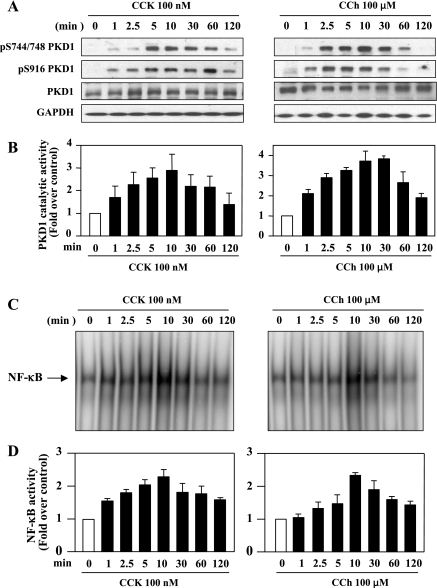
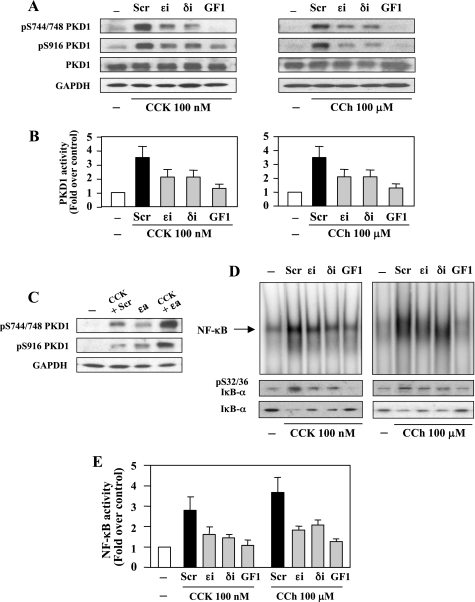
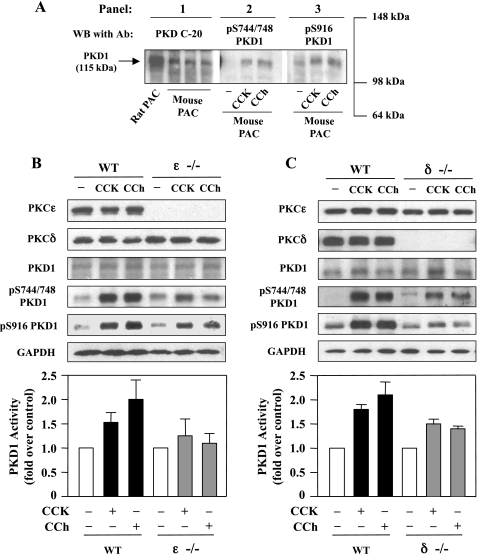
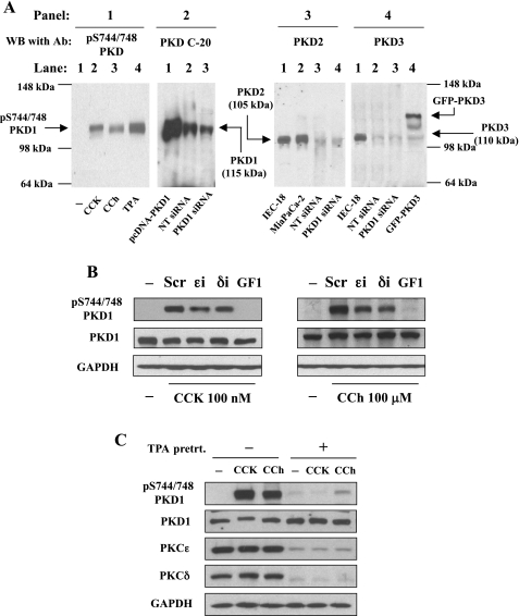
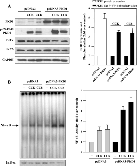
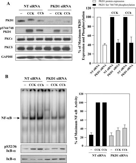
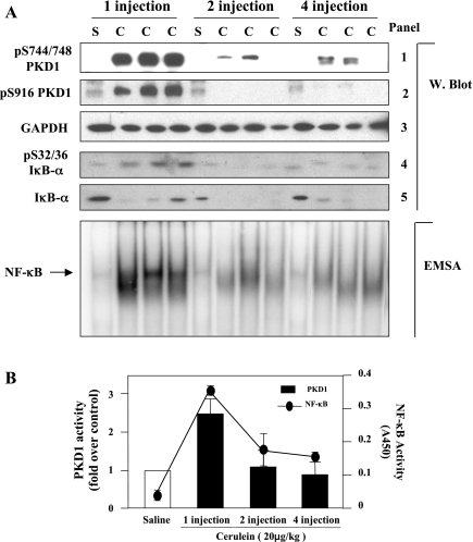
The transcription factor NF-kappaB plays a critical role in inflammatory and cell death responses during acute pancreatitis. Previous studies in our laboratory demonstrated that protein kinase C (PKC) isoforms PKCdelta and epsilon are key regulators of NF-kappaB activation induced by cholecystokinin-8 (CCK-8), tumor necrosis factor-alpha, and ethanol. However, the downstream participants in regulating NF-kappaB activation in exocrine pancreas remain poorly understood. Here, we demonstrate that protein kinase D1 (PKD1) is a key downstream target of PKCdelta and PKCepsilon in pancreatic acinar cells stimulated by two major secretagogues, CCK-8 and the cholinergic agonist carbachol (CCh), and that PKD1 is necessary for NF-kappaB activation induced by CCK-8 and CCh. Both CCK-8 and CCh dose dependently induced a rapid and striking activation of PKD1 in rat pancreatic acinar cells, as measured by in vitro kinase assay and by phosphorylation at PKD1 activation loop (Ser744/748) or autophosphorylation site (Ser916). The phosphorylation and activation of PKD1 correlated with NF-kappaB activity stimulated by CCK-8 or CCh, as measured by NF-kappaB DNA binding. Either inhibition of PKCdelta or epsilon by isoform-specific inhibitory peptides, genetic deletion of PKCdelta and epsilon in pancreatic acinar cells, or knockdown of PKD1 by using small interfering RNAs in AR42J cells resulted in a marked decrease in PKD1 and NF-kappaB activation stimulated by CCK-8 or CCh. Conversely, overexpression of PKD1 resulted in augmentation of CCK-8- and CCh-stimulated NF-kappaB activation. Finally, the kinetics of PKD1 and NF-kappaB activation during cerulein-induced rat pancreatitis showed that both PKD1 and NF-kappaB activation were early events during acute pancreatitis and that their time courses of response were similar. Our results identify PKD1 as a novel early convergent point for PKCdelta and epsilon in the signaling pathways mediating NF-kappaB activation in pancreatitis.


Figures

Fig. 1.
Fig. 1.
CCK-8 and carbachol (CCh) induce PKD1 phosphorylation and activation and NF-κB activation in rat pancreatic acinar cells. Rat pancreatic acinar cells were incubated with 100 nM CCK-8 (CCK) or 100 μM CCh for the indicated time periods. A: Western blotting analyses of the time course of CCK8- and CCh-induced PKD1 phosphorylation by use of PKD1 pS744/748 antibody or PKD1 pS916 antibody. Blots were reblotted for PKD1 expression with PKD C-20 antibody, and then for GAPDH to verify equal protein loading. All Western blots shown are representative of 3 independent experiments. B: time course of CCK8- and CCh-induced PKD-1 catalytic activation. The acinar lysates were immunoprecipitated using PKD C-20 antibody, and PKD1 activity in the immunocomplexes was determined by in vitro kinase assay. Results are expressed as a fold increase over unstimulated control in activity. Values are means ± SE (n = 3). C: time course of CCK-8 and CCh-induced NF-κB binding activity measured by EMSA. D: NF-κB band intensities were quantified in the PhosphorImager and normalized on the band intensity in unstimulated control. Values are means ± SE (n = 3).
Fig. 2.
Fig. 2.
PKC inhibitors prevent CCK-8- and CCh-stimulated PKD1 and NF-κB activation in pancreatic acinar cells. Rat pancreatic acinar cells were incubated for 3 h with PKCɛ translocation inhibitor (ɛi), PKC-δ translocation inhibitor (δi), or scrambled peptide (Scr) (10 μM each), or with the broad-spectrum PKC inhibitor GF1 (3.5 μM) before stimulation with 100 nM CCK-8 or 100 μM CCh for 10 min. A: cell lysates were analyzed by Western blot using PKD1 pS744/748 antibody or PKD1 pS916 antibody. The blots were reprobed for PKD1 then for GAPDH to verify equal protein loading. B: the acinar cell lysates were immunoprecipitated with PKD C-20 antibody, and PKD1 catalytic activity in the immunocomplexes was determined by in vitro kinase assay. The results are expressed as a fold increase over unstimulated control in activity. Values are means ± SE (n = 3). C: rat pancreatic acinar cells were incubated for 3 h with PKCɛ translocation activator (ɛa) or scrambled peptide (10 μM each), and 0.1 nM CCK was added during the last 10 min of the incubation. Cell lysates were analyzed by Western blot using PKD1 pS744/748 antibody or PKD1 pS916 antibody. The blots were reprobed for GAPDH to verify equal protein loading. D: NF-κB binding activity was measured in nuclear extracts by EMSA. IκΒ-α phosphorylation and degradation were measured in cytosolic extracts from the same samples by Western blot analysis. E: NF-κB band intensities were quantified in the PhosphorImager and normalized on the band intensity in unstimulated control acini. Values are means ± SE (n = 3).
Fig. 3.
Fig. 3.
PKD1 phosphorylation and activation induced by either CCK-8 or CCh is reduced in mice deficient in PKCɛ or PKCδ. A: mouse pancreatic acinar cells (PAC) were incubated for 10 min with 100 nM CCK-8 or with 100 μM CCh and the cell lysates were analyzed by Western blotting using antibodies against PKD C-20 (panel 1), pS744/748 PKD1 (panel 2), or pS916 PKD1 (panel 3). B and C: pancreatic acinar cells isolated from wild-type (WT) mice and mice deficient in PKCɛ (ɛ−/−) and PKCδ (δ−/−) were incubated with 100 nM CCK-8 or 100 μM CCh for 10 min at 37°C and the cell lysates were analyzed by Western blot with antibodies against PKCɛ, PKCδ, PKD1 C-20, pS744/748 PKD1, or pS916 PKD1. The blots were then reprobed for GAPDH to verify equal protein loading. Bar graphs: mouse acinar cell lysates were immunoprecipitated with PKD C-20 antibody and PKD1 catalytic activity in the immunocomplexes was determined by in vitro kinase assay. The results are expressed as a fold increase over unstimulated control in activity. Values are means ± SE (n = 2). Representative of 2 independent experiments.
Fig. 4.
Fig. 4.
PKD1 is the predominant PKD isoform in AR42J cells and is activated by CCK-8 and CCh through a PKC-dependent pathway. A, panel 1: Western blot analysis using anti-pS744/748 PKD antibody for the lysate samples from AR42J cells incubated for 10 min at 37° with 10 nM CCK-8 or 100 μM CCh. Panel 2: lysates from AR42J cells transfected with pcDNA3-PKD1, nontargeting siRNAs (NT), or PKD1 siRNAs were analyzed by Western blot with PKD C-20 antibody that recognizes the COOH-terminal region of both PKD1 and PKD2. Panel 3: Western blot with a specific PKD2 antibody for the lysate samples from rat IEC-18, MiaPaCa-2, AR42J transfected with nontargeting siRNAs, or PKD1 siRNAs. Panel 4: Western blot with a specific PKD3 antibody for lysates from AR42J cells transfected with nontargeting siRNAs or PKD1 siRNAs using the lysates from IEC-18 cells and AR42J cells transfected with GFP-PKD3 as positive controls of PKD3. All Western blot results are representatives of 3 experiments. B: AR42J cells were incubated for 3 h with PKCɛ translocation inhibitor, PKCδ translocation inhibitor (δi), or scrambled peptide, 10 μM each, or with 3.5 μM broad-spectrum PKC inhibitor GF1. Then 100 nM CCK-8 or 100 μM CCh was added during the last 10 min of the incubation. Cell lysates were analyzed by Western blot using PKD1 pS744/748 antibody. The blots were reprobed for PKD1 expression by using PKD C-20 antibody and then for GAPDH to verify equal protein loading. C: serum-starved cultures of AR42J cells were incubated for 40 h either without (−) or with (+) 1 μM 12-O-tetradecanoylphorbol-13-acetate (TPA). The cells were washed and incubated for 4 h at 37°C in serum-free medium. Cells were then incubated for a further 10 min without or with CCK-8 (100 nM) or CCh (100 μM). Cell lysates were analyzed by Western blot with antibodies against pS744/748 PKD1, PKD1 C-20, PKCɛ, and PKCδ. To verify equal protein loading, the blots were reblotted for GAPDH.
Fig. 5.
Fig. 5.
Overexpression of PKD1 enhances NF-κB activation induced by CCK-8 or CCh. AR42J cells were transfected with the empty vector pcDNA3 or pcDNA3-PKD1. Two days after transfection, the cells were washed and incubated in serum-free F-12K medium for 3 h. Then the cells were incubated for 10 min in the absence (−) or presence (+) of 100 nM CCK-8 or 100 μM CCh. A, left: Western blot with antibodies against PKD C-20 for PKD1 expression or against pS744/748 for PKD1 phosphorylation. Western blots were also probed for the expression of PKCɛ and δ and then further probed for GAPDH to verify equal protein loading. Shown are representative blots from at least 3 independent experiments. Right: intensity of the Western blot bands was quantified by densitometry for the level of PKD1 expression (open bars) and PKD1 Ser744/748 phosphorylation (solid bars), expressed as fold increase relative to the controls (pcDNA3-transfected cells). Values are means ± SE (n = 6). B, left: NF-κB DNA binding activity in nuclear extracts was measured by EMSA, and IκΒ-α degradation in cytosolic extracts were measured by Western blot analysis. Right: NF-κB band intensities were quantified in the PhosphorImager and normalized on the band intensity in unstimulated control acinar cells. Values are means ± SE (n = 3).
Fig. 6.
Fig. 6.
PKD1 knockdown decreases NF-κB activation induced by CCK-8 and CCh. AR42J cells were transiently transfected with PKD1 siRNAs or nontargeting negative control siRNAs. Two days after transfection, the cells were washed and incubated in serum-free F-12K medium for 3 h. Then the cells were incubated for 10 min in the absence or presence of 100 nM CCK-8 or 100 μM CCh. A, left: Western blot with antibodies against PKD1 C-20 for PKD1 expression or against pS744/748 for PKD1 phosphorylation. Western blots were also probed for the expression of PKCɛ and δ and then further probed for GAPDH to verify equal protein loading. Shown are representative blots from at least 3 independent experiments. Right: the intensity of the Western blot bands was quantified by densitometry for the level of PKD1 expression (open bars) and PKD1 Ser744/748 phosphorylation (solid bars), expressed as % of maximum expression level of PKD1 (open bars) or % of maximum in PKD1 phosphorylation obtained with 100 nM CCK-8 or 100 μM CCh. The PKD1 expression level or Ser744/748 phosphorylation in NT siRNAs control were considered as 100%. Values are means ± SE (n = 6). B, left: NF-κB DNA binding activity in nuclear extracts was measured by EMSA; IκΒ-α phosphorylation and degradation in cytosolic extracts were measured by Western blot analysis. Right: NF-κB band intensities were quantified in the PhosphorImager and normalized on the band intensity in unstimulated control acinar cells. Values are means ± SE (n = 3).
Fig. 7.
Fig. 7.
PKD1 and NF-κB are time dependently activated by cerulein in rat cerulein pancreatitis. Pancreatitis was induced in rats by 4 hourly intraperitoneal injections of cerulein (C, 20 μg/kg); control animals received similar injections of saline (S). Animals were then euthanized in 30 min after the 1st, 2nd, and 4th injection. Induction of pancreatitis was confirmed by elevated levels of serum amylase and lipase and by changes in pancreatic histology on tissue sections. A: time course of PKD1 phosphorylation and NF-κB activation in rat cerulein pancreatitis. Western blot: tissue lysates were analyzed by Western blot using PKD1 pS744/748 antibody (panel 1) or PKD1 pS916 antibody (panel 2). The blots were then reblotted with GAPDH antibody to verify equal protein loading (panel 3). IκΒ-α phosphorylation and degradation in cytosolic extracts were measured by Western blot analysis (panels 4 and 5). EMSA: time course of NF-κB activation in rat cerulein pancreatitis. EMSA was performed on nuclear extracts from the rat pancreas tissue. B: time course of PKD1 kinase activity and NF-κB activation in rat cerulein pancreatitis. The tissue lysates were immunoprecipitated with PKD1 C-20 antibody and PKD1 activity in the immunocomplexes was determined by in vitro kinase assay. The results (bar graphs) are expressed as a fold increase over saline control in activity. Values are means ± SE (n = 3). NF-κB binding activity of the nuclear extracts was determined by ELISA using ActiveMotif kit. Values presented in the curve figure are means ± SE (n = 3).

Similar articles

Cited by

References

    1. Adler G, Beglinger C, Braun U, Reinshagen M, Koop I, Schafmayer A, Rovati L, Arnold R. Interaction of the cholinergic system and cholecystokinin in the regulation of endogenous and exogenous stimulation of pancreatic secretion in humans. Gastroenterology 100: 537–543, 1991. - PubMed
    1. Akiyama T, Hirohata Y, Okabayashi Y, Imoto I, Otsuki M. Supramaximal CCK and CCh concentrations abolish VIP potentiation by inhibiting adenylyl cyclase activity. Am J Physiol Gastrointest Liver Physiol 275: G1202–G1208, 1998. - PubMed
    1. Bastani B, Yang L, Baldassare JJ, Pollo DA, Gardner JD. Cellular distribution of isoforms of protein kinase C (PKC) in pancreatic acini. Biochim Biophys Acta 1269: 307–315, 1995. - PubMed
    1. Berna MJ, Hoffmann KM, Tapia JA, Thill M, Pace A, Mantey SA, Jensen RT. CCK causes PKD1 activation in pancreatic acini by signaling through PKC-delta and PKC-independent pathways. Biochim Biophys Acta 1773: 483–501, 2007. - PMC - PubMed
    1. Chen X, Ji B, Han B, Ernst SA, Simeone D, Logsdon CD. NF-kappaB activation in pancreas induces pancreatic and systemic inflammatory response. Gastroenterology 122: 448–457, 2002. - PubMed

Publication types

MeSH terms