

Expression of peroxisome proliferator-activated receptor-gamma in key neuronal subsets regulating glucose metabolism and energy homeostasis
- PMID: 18845632
- PMCID: PMC2646542
- DOI: 10.1210/en.2008-0899
Expression of peroxisome proliferator-activated receptor-gamma in key neuronal subsets regulating glucose metabolism and energy homeostasis
Abstract
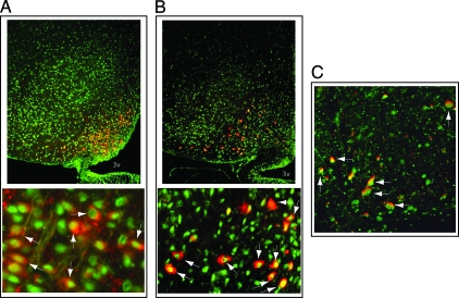
In addition to increasing insulin sensitivity and adipogenesis, peroxisome proliferator-activated receptor (PPAR)-gamma agonists cause weight gain and hyperphagia. Given the central role of the brain in the control of energy homeostasis, we sought to determine whether PPARgamma is expressed in key brain areas involved in metabolic regulation. Using immunohistochemistry, PPARgamma distribution and its colocalization with neuron-specific protein markers were investigated in rat and mouse brain sections spanning the hypothalamus, the ventral tegmental area, and the nucleus tractus solitarius. In several brain areas, nuclear PPARgamma immunoreactivity was detected in cells that costained for neuronal nuclei, a neuronal marker. In the hypothalamus, PPARgamma immunoreactivity was observed in a majority of neurons in the arcuate (including both agouti related protein and alpha-MSH containing cells) and ventromedial hypothalamic nuclei and was also present in the hypothalamic paraventricular nucleus, the lateral hypothalamic area, and tyrosine hydroxylase-containing neurons in the ventral tegmental area but was not expressed in the nucleus tractus solitarius. To validate and extend these histochemical findings, we generated mice with neuron-specific PPARgamma deletion using nestin cre-LoxP technology. Compared with littermate controls, neuron-specific PPARgamma knockout mice exhibited dramatic reductions of both hypothalamic PPARgamma mRNA levels and PPARgamma immunoreactivity but showed no differences in food intake or body weight over a 4-wk study period. We conclude that: 1) PPARgamma mRNA and protein are expressed in the hypothalamus, 2) neurons are the predominant source of PPARgamma in the central nervous system, although it is likely expressed by nonneuronal cell types as well, and 3) arcuate nucleus neurons that control energy homeostasis and glucose metabolism are among those in which PPARgamma is expressed.
Figures


References
-
- Rosen ED, Sarraf P, Troy AE, Bradwin G, Moore K, Milstone DS, Spiegelman BM, Mortensen RM 1999 PPARγ is required for the differentiation of adipose tissue in vivo and in vitro. Mol Cell 4:611–617 - PubMed
-
- Spiegelman BM 1998 PPAR-γ: adipogenic regulator and thiazolidinedione receptor. Diabetes 47:507–514 - PubMed
-
- Escher P, Braissant O, Basu-Modak S, Michalik L, Wahli W, Desvergne B 2001 Rat PPARs: quantitative analysis in adult rat tissues and regulation in fasting and refeeding. Endocrinology 142:4195–4202 - PubMed
-
- Rosen ED, Kulkarni RN, Sarraf P, Ozcan U, Okada T, Hsu CH, Eisenman D, Magnuson MA, Gonzalez FJ, Kahn CR, Spiegelman BM 2003 Targeted elimination of peroxisome proliferator-activated receptor γ in β cells leads to abnormalities in islet mass without compromising glucose homeostasis. Mol Cell Biol 23:7222–7229 - PMC - PubMed
-
- Hevener AL, Olefsky JM, Reichart D, Nguyen MT, Bandyopadyhay G, Leung HY, Watt MJ, Benner C, Febbraio MA, Nguyen AK, Folian B, Subramaniam S, Gonzalez FJ, Glass CK, Ricote M 2007 Macrophage PPARγ is required for normal skeletal muscle and hepatic insulin sensitivity and full antidiabetic effects of thiazolidinediones. J Clin Invest 117:1658–1669 - PMC - PubMed
Publication types
MeSH terms
Substances
Grants and funding
- DK064857/DK/NIDDK NIH HHS/United States
- R01 DK052989/DK/NIDDK NIH HHS/United States
- R01 DK069927/DK/NIDDK NIH HHS/United States
- DK52989/DK/NIDDK NIH HHS/United States
- P30 DK035816/DK/NIDDK NIH HHS/United States
- DK20593/DK/NIDDK NIH HHS/United States
- R01 NS032273/NS/NINDS NIH HHS/United States
- DK59637/DK/NIDDK NIH HHS/United States
- P30 DK020593/DK/NIDDK NIH HHS/United States
- DK68340/DK/NIDDK NIH HHS/United States
- K08 DK064857/DK/NIDDK NIH HHS/United States
- NS32273/NS/NINDS NIH HHS/United States
- P30 DK-17047/DK/NIDDK NIH HHS/United States
- DK069927/DK/NIDDK NIH HHS/United States
- U24 DK059637/DK/NIDDK NIH HHS/United States
- P30 DK017047/DK/NIDDK NIH HHS/United States
LinkOut - more resources
Full Text Sources
Molecular Biology Databases
Research Materials
