Comparative Study
. 2009 Feb;150(2):651-61.
doi: 10.1210/en.2008-1118. Epub 2008 Oct 9.

Lean phenotype and resistance to diet-induced obesity in vitamin D receptor knockout mice correlates with induction of uncoupling protein-1 in white adipose tissue

Affiliations
Comparative Study

Lean phenotype and resistance to diet-induced obesity in vitamin D receptor knockout mice correlates with induction of uncoupling protein-1 in white adipose tissue

Carmen J Narvaez et al. Endocrinology. 2009 Feb.

Abstract

Increased adiposity is a feature of aging in both mice and humans, but the molecular mechanisms underlying age-related changes in adipose tissue stores remain unclear. In previous studies, we noted that 18-month-old normocalcemic vitamin D receptor (VDR) knockout (VDRKO) mice exhibited atrophy of the mammary adipose compartment relative to wild-type (WT) littermates, suggesting a role for VDR in adiposity. Here we monitored body fat depots, food intake, metabolic factors, and gene expression in WT and VDRKO mice on the C57BL6 and CD1 genetic backgrounds. Regardless of genetic background, both sc and visceral white adipose tissue depots were smaller in VDRKO mice than WT mice. The lean phenotype of VDRKO mice was associated with reduced serum leptin and compensatory increased food intake. Similar effects on adipose tissue, leptin and food intake were observed in mice lacking Cyp27b1, the 1alpha-hydroxylase enzyme that generates 1,25-dihydroxyvitamin D(3), the VDR ligand. Although VDR ablation did not reduce expression of peroxisome proliferator-activated receptor-gamma or fatty acid synthase, PCR array screening identified several differentially expressed genes in white adipose tissue from WT and VDRKO mice. Uncoupling protein-1, which mediates dissociation of cellular respiration from energy production, was greater than 25-fold elevated in VDRKO white adipose tissue. Consistent with elevation in uncoupling protein-1, VDRKO mice were resistant to high-fat diet-induced weight gain. Collectively, these studies identify a novel role for 1,25-dihydroxyvitamin D(3) and the VDR in the control of adipocyte metabolism and lipid storage in vivo.


Figures

Figure 1
Figure 1
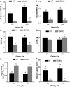
Adipose stores and food intake in WT and VDRKO mice. A, Representative images of visceral adipose stores in 18-month WT (left panel) and VDRKO (right panel) mice fed the high-calcium rescue diet from weaning. Body weights (B) and abdominal WAT (C) were measured in normocalcemic WT and VDRKO mice on the C57BL6 background at 2, 4, and 6 months of age. D, Serum leptin was measured by ELISA. E, Individual food intake was measured over the 10-d period preceding the time the animals were killed. All data are mean ± se. *, P < 0.05, WT vs. VDRKO.
Figure 2
Figure 2
Effects of high-fat diet on tissue weights, food intake, and serum leptin in WT and VDRKO mice on the CD1 genetic background. WT and VDRKO mice on the CD1 background were weaned onto the high-calcium rescue diet containing either 5 or 15% fat from corn oil. Diets were provided ad libitum and mice were killed at 6 months of age. A, Abdominal WAT. B, inguinal WAT. C, iBAT. D, Liver. E, Caloric intake was measured in individually caged mice for the 10 d preceding the time the animals were killed. F, Serum leptin was assessed by ELISA. All data are mean ± se; bars with different letters (a–d) are significantly different by ANOVA (P < 0.05).
Figure 3
Figure 3
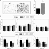
WAT histology and metabolic parameters in WT and VDRKO mice. A. Left panel, Representative H&E-stained sections of abdominal WAT from 6-month-old WT and VDRKO mice on the C57BL6 background. Fat pads were collected, fixed, and subjected to H&E staining. A representative image is shown for each fat pad/genotype. Right panel, The number of cells per field was counted for three to four mice per genotype on the CD1 background; bars represent mean ± se. *, P < 0.05 by Student’s t test. Serum was collected from C57BL6 (B) and CD1 (C) mice for analysis of glucose, insulin, and triglycerides as described in Materials and Methods. Background lines represent the reference values (mean ± se) for C57BL6 mice (obtained from the Mouse Phenome Database at www.jax.org) for each parameter. Data bars are mean ± se; bars with different letters (a–c) are significantly different by ANOVA (P < 0.05).
Figure 4
Figure 4
Analysis of WT and Cyp27b1 knockout (KO) mice. Top panel, Female WT and Cyp27b1 KO mice on the C57BL6 background were fed a high-calcium rescue diet containing 15% fat from weaning and body weights (BW) were monitored through 6 months of age. Data are mean values of four to seven mice. *, P < 0.05, WT vs. Cyp27b1 knockout. Bottom panel, Tissue weights, serum leptin, and food intake of WT and Cyp27b1 knockout mice on the standard rescue diet were measured at 9 months of age as described in Materials and Methods. Data represent mean ± se. *, P < 0.05, WT vs. Cyp27b1 knockout.
Figure 5
Figure 5
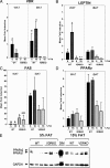
Effect of VDR genotype on gene and protein expression in white and brown adipose tissue. Gene expression was determined by real-time PCR in WAT and iBAT from normocalcemic 6-month-old WT mice on the CD1 background fed two levels of dietary fat (5 vs. 15%) from weaning. Data were calculated by the comparative cycle threshold method, normalized against 18S and expressed relative to values from WAT of WT mice fed 5% fat, which was set at 1. A, VDR. B, Leptin. C, FAS. D, PPARγ. For A–D, bars represent mean ± se. *, Significantly different (P < 0.05) compared with WT mice fed 5% fat. Bars with different letters are significantly different by ANOVA (P < 0.05). E, Western blot of PPARγ protein expression in WAT from two to three individual mice per group. Both PPARγ1 and PPARγ2 isoforms were detected in WAT, but no consistent differences were observed by diet or genotype. DIM, Cells treated with adipogenic cocktail used as positive control.
Figure 6
Figure 6
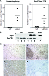
Effect of VDR ablation on ucp-1 expression in WAT. A, Relative ucp-1 gene expression normalized against hypoxanthine-guanine phosphoribosyltransferase (calculated from screening array data) in abdominal WAT of 6-month-old CD1 WT and VDRKO mice fed the high-calcium rescue diet containing 5% fat. Each symbol represents tissue from an individual mouse, and horizontal bar represents group mean. B, Confirmation of ucp-1 up-regulation in WAT from 6-month-old VDRKO mice by real-time PCR. Each symbol represents tissue from an individual WT or VDRKO CD1 mouse fed the high-calcium rescue diet containing 5% fat; horizontal bar represents group mean. C, Western blot of ucp-1 protein expression in WAT (50 μg/lane) from three individual mice per group (6 month old WT or VDRKO mice fed 5% fat). Lanes 1–3, WT; lanes 4–6, VDRKO; lane 7, iBAT lysate (2 μg/lane) was used as positive control for ucp-1. Blots were stripped and reprobed with anti-GAPDH as loading control. D, Immunohistochemistry of ucp-1 in formalin-fixed sections of WAT from 6-month-old CD1 WT (A) and VDRKO (B) mice fed 5% fat. Sections were incubated with antibody against ucp-1 (5 μg/ml) and developed with the diaminobenzidine procedure. Brown adipose tissue was used as positive control (C) and no antibody negative control (D). Slides were counterstained with hematoxylin.

References

    1. Imai T, Jiang M, Chambon P, Metzger D 2001 Impaired adipogenesis and lipolysis in the mouse upon selective ablation of the retinoid X receptor α mediated by a tamoxifen-inducible chimeric Cre recombinase (Cre-ERT2) in adipocytes. Proc Natl Acad Sci USA 98:224–228 - PMC - PubMed
    1. Kalaany NY, Gauthier KC, Zavacki AM, Mammen PP, Kitazume T, Peterson JA, Horton JD, Garry DJ, Bianco AC, Mangelsdorf DJ 2005 LXRs regulate the balance between fat storage and oxidation. Cell Metab 1:231–244 - PubMed
    1. Sjögren M, Alkemade A, Mittag J, Nordström K, Katz A, Rozell B, Westerblad H, Arner A, Vennström B 2007 Hypermetabolism in mice caused by the central action of an unliganded thyroid hormone receptor α1. EMBO J 26:4535–4545 - PMC - PubMed
    1. Kamei Y, Kawada T, Kazuki R, Ono T, Kato S, Sugimoto E 1993 Vitamin D receptor gene expression is up-regulated by 1, 25-dihydroxyvitamin D3 in 3T3-L1 preadipocytes. Biochem Biophys Res Commun 193:948–955 - PubMed
    1. Imagawa M, Tsuchiya T, Nishihara T 1999 Identification of inducible genes at the early stage of adipocyte differentiation of 3T3-L1 cells. Biochem Biophys Res Commun 254:299–305 - PubMed

Publication types

MeSH terms