Comparative Study
. 2009 Feb;150(2):849-61.
doi: 10.1210/en.2008-1069. Epub 2008 Oct 9.

Endothelial dysfunction in mice with streptozotocin-induced type 1 diabetes is opposed by compensatory overexpression of cyclooxygenase-2 in the vasculature

Affiliations
Comparative Study

Endothelial dysfunction in mice with streptozotocin-induced type 1 diabetes is opposed by compensatory overexpression of cyclooxygenase-2 in the vasculature

Carmela Nacci et al. Endocrinology. 2009 Feb.

Abstract

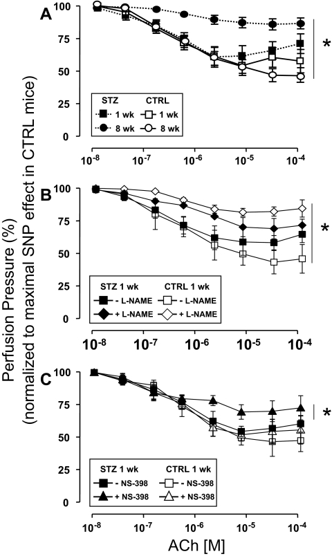
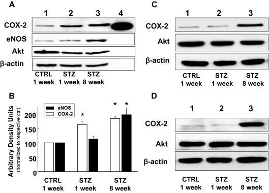
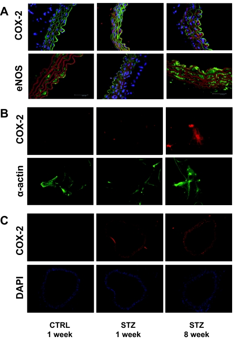
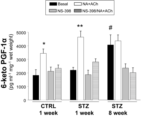
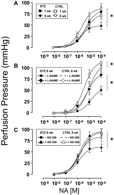
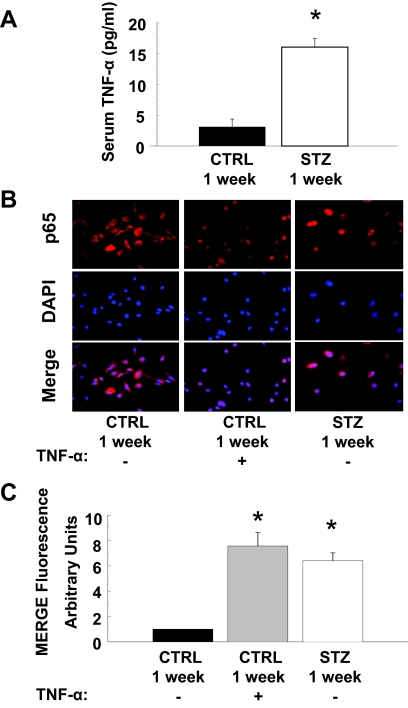
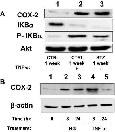
Cardiovascular complications of diabetes result from endothelial dysfunction secondary to persistent hyperglycemia. We investigated potential compensatory mechanisms in the vasculature that oppose endothelial dysfunction in diabetes. BALB/c mice were treated with streptozotocin (STZ) to induce type 1 diabetes (T1D). In mesenteric vascular beds (MVBs), isolated ex vivo from mice treated with STZ for 1 wk, dose-dependent vasorelaxation to acetylcholine (ACh) or sodium nitroprusside was comparable with that in age-matched control mice (CTRL). By contrast, MVBs from mice treated with STZ for 8 wk had severely impaired vasodilator responses to ACh consistent with endothelial dysfunction. Pretreatment of MVBs from CTRL mice with nitric oxide synthase inhibitor nearly abolished vasodilation to ACh. In MVB from 1-wk STZ-treated mice, vasodilation to ACh was only partially impaired by L-N(omega)-arginine methyl ester. Thus, vasculature of mice with T1D may have compensatory nitric oxide-independent mechanisms to augment vasodilation to ACh and oppose endothelial dysfunction. Indeed, pretreatment of MVBs isolated from 1-wk STZ-treated mice with NS-398 [selective cyclooxygenase (COX)-2 inhibitor] unmasked endothelial dysfunction not evident in CTRL mice pretreated without or with NS-398. Expression of COX-2 in MVBs, aortic endothelial cells, and aortic vascular smooth muscle cells from STZ-treated mice was significantly increased (vs. CTRL). Moreover, concentrations of the COX-2-dependent vasodilator 6-keto-prostaglandin F-1alpha was elevated in conditioned media from aorta of STZ-treated mice. We conclude that endothelial dysfunction in a mouse model of T1D is opposed by compensatory up-regulation of COX-2 expression and activity in the vasculature that may be relevant to developing novel therapeutic strategies for diabetes and its cardiovascular complications.


Figures

Figure 1
Figure 1
Endothelial dysfunction in STZ-induced diabetes is unmasked by inhibition of COX-2. MVBs were isolated from mice treated with STZ (closed symbols) or vehicle CTRL (open symbols) and prepared as described in Materials and Methods. For these experiments, MVBs were contracted to about 80% of maximal vasoconstriction. To obtain comparable levels of precontraction with a perfusion pressure of about 100 mm Hg in MVBs from CTRL and STZ mice, 3 μm NA were used in MVBs from 1-wk STZ and 1-wk CTRL mice. A, Dose-response curves for ACh-induced vasorelaxation were obtained in MVBs from mice 1 wk (squares) or 8 wk (circles) after treatment with STZ or vehicle control. Results are mean ± sem of 11 (STZ) and eight (CTRL) independent experiments. ACh-mediated vasorelaxation was significantly impaired in MVBs from mice treated with STZ for 8 wk (vs. respective CTRL, P < 0.001). No significant difference was observed for ACh-induced vasodilation when results from 1-wk STZ and 8-wk CTRL were compared (P > 0.09). B, Dose-response curves for ACh-mediated vasorelaxation were obtained in MVBs from mice 1 wk after treatment with STZ or vehicle control in the absence (squares) or presence (diamonds) of pretreatment with the NO synthase antagonist L-NAME (100 μm, 30 min). Pretreatment with L-NAME significantly increased perfusion pressure to a similar extent (∼10 mm Hg) in MVBs from both CTRL and STZ-treated mice. Results are mean ± sem of five (STZ) and four (CTRL) independent experiments. Inhibition of NO synthase significantly reduced ACh-mediated vasorelaxation in MVBs from CTRL (vs. respective basal, P < 0.001) but not 1-wk STZ-treated mice. A statistical difference was observed in ACh-induced vasodilation when results from 1-wk CTRL + L-NAME and 1-wk STZ + L-NAME were compared (P < 0.05); no significant difference was observed in ACh-induced vasodilation when results from 1-wk CTRL without L-NAME and 1-wk STZ without L-NAME were compared (P > 0.12). C, Dose-response curves for ACh-mediated vasorelaxation were obtained in MVBs from mice 1 wk after treatment with STZ or vehicle control in the absence (squares) or presence (triangles) of pretreatment with the specific COX-2 inhibitor NS-398 (10 μm, 30 min). Results are mean ± sem of seven (STZ) and four (CTRL) independent experiments. ACh-mediated vasorelaxation was significantly impaired in MVBs from mice treated with STZ for 1 wk in the presence of NS-398 pretreatment (when compared with MVBs from 1 wk STZ in the absence of NS-398, P < 0.02). No significant difference was observed in ACh-induced vasodilation when results from 1-wk CTRL without NS-398 and 1-wk STZ without NS-398 were compared (P > 0.14). Asterisks refer to significant differences found between indicated curves assessed by two-way ANOVA for repeated measures.
Figure 2
Figure 2
Expression of COX-2 and eNOS, but not Akt, is elevated in vasculature of mice with STZ-induced Diabetes. A, Lysates of MVBs obtained from mice treated with STZ or CTRL for the indicated times were immunoblotted with indicated antibodies. Time-dependent increase in COX-2 and eNOS expression in MVBs after STZ treatment in a representative immunoblot from three independent experiments. Lane 4, COX-2 purified protein, positive control. B, Mean ± sem of densitometric analysis for COX-2 and eNOS expression normalized for Akt expression for three independent experiments. Significant increases in protein expression were observed for COX-2 in MVBs of mice treated with STZ for 1 wk and COX-2 and eNOS in MVBs of mice treated with STZ for 8 wk (vs. CTRL, P < 0.05). Asterisks refer to significant differences found between indicated groups assessed by Student’s t test. C, Lysates from aortas of mice treated with vehicle control or STZ for the indicated times were immunoblotted with indicated antibodies. Representative immunoblots of experiments were repeated independently three times. D, Lysates of VSMCs from aortas of mice treated with vehicle control or STZ for the indicated times were immunoblotted with indicated antibodies. Representative immunoblots from experiments independently repeated three times are shown.
Figure 3
Figure 3
Expression of COX-2 and eNOS is increased in aortas from mice with STZ-induced diabetes. Aortas from mice treated with CTRL or STZ for the indicated times were prepared as described in Materials and Methods. A, Aortas were fixed in 8% paraformaldehyde and ring sections (10 μm) were immunolabeled with antibodies against COX-2 (upper panel) or eNOS (lower panel). Sections were immunostained with IgG-fluorescein isothiocyanate conjugated- (upper panel) and IgG-Texas Red conjugated (lower panel)-laminin antibodies. Finally, DAPI staining was used to visualize cell nuclei. Thus, visualization by confocal microscopy shows red fluorescence for COX-2 staining and green fluorescence for laminin staining in the upper panel. In the lower panel, green fluorescence is seen for eNOS staining, whereas red fluorescence is seen for laminin staining. Cell nuclei are visualized in blue. Representative results are shown for experiments independently repeated three times. B, VSMCs were prepared from aortas of mice treated with CTRL or STZ for the indicated times as described in Materials and Methods. VSMCs were fixed in 3.5% paraformaldehyde and immunolabeled with antibodies against COX-2 (upper panel) or α-actin (lower panel). Representative results are shown for experiments that were repeated independently three times. C, Mesenteric arteries were fixed in 8% paraformaldehyde and ring sections (8 μm) were immunolabeled with COX-2 antibody (upper panel). DAPI staining was used to visualize cell nuclei (lower panel). Red fluorescence indicates COX-2 staining. Cell nuclei are visualized in blue. Representative results are shown for experiments independently repeated three times.
Figure 4
Figure 4
Production of 6-keto-PGF-1α is elevated in aortas of STZ-treated mice. Aortas from mice treated with CTRL or STZ for the indicated times were prepared as described in Materials and Methods. Aortas were incubated in Krebs-Henseleit solution (20 min, 37 C) and then treated without or with NA + ACh in the absence or presence of COX-2 inhibitor NS-398 (10 μm). Conditioned media were then collected for measurement of 6-keto-PGF-1α (ELISA). Levels of 6-keto-PGF-1α were significantly increased in response to NA + ACh stimulation in conditioned media from cultured aortas isolated from mice 1 wk after injection with CTRL or STZ (vs. respective basal). *, P < 0.005; **, P < 0.001. Significant elevations of 6-keto-PGF-1α concentrations in conditioned media were observed both before and after treatment with NA + ACh in aortas isolated from mice 8 wk after injection with STZ (vs. CTRL basal. #, P < 0.01. Results are mean ± sem (normalized to wet weight of aorta) for experiments repeated independently three times in duplicate.
Figure 5
Figure 5
Both COX-2- and NO-dependent mechanisms participate in opposing endothelial dysfunction in STZ-induced diabetes. MVBs were isolated from mice treated with STZ (closed symbols) or vehicle control (open symbols) and prepared as described in Materials and Methods. A, Dose-response curves for NA-induced vasoconstriction were obtained in MVBs from mice 1 wk (squares) or 8 wk (circles) after treatment with STZ or vehicle control. Results are mean ± sem of 12 (STZ) and 10 (CTRL) independent experiments. NA-mediated vasoconstriction was significantly impaired in MVB from mice treated with STZ for 8 wk (vs. respective CTRL, P < 0.001). B, Dose-response curves for NA-mediated vasoconstriction were obtained in MVBs from mice 8 wk after treatment with STZ or vehicle control in the absence (circles) or presence (diamonds) of pretreatment with the NO synthase antagonist L-NAME (100 μm, 30 min). Results are mean ± sem of four (STZ) and five (CTRL) independent experiments. Inhibition of NO synthase significantly increased NA-mediated vasoconstriction in MVBs from mice treated with either STZ or vehicle control for 8 wk (vs. results without L-NAME pretreatment, P < 0.01). C, Dose-response curves for NA-mediated vasoconstriction were obtained in MVB from mice 8 wk after treatment with STZ or vehicle control in the absence (circles) or presence (triangles) of pretreatment with the specific COX-2 inhibitor NS-398 (10 μm, 30 min). Results are mean ± sem of eight (STZ) and seven (CTRL) independent experiments. Inhibition of COX-2 significantly increased NA-mediated vasoconstriction in MVB from mice treated with STZ for 8 wk (vs. results without NS-398 pretreatment, P < 0.01) but not in MVB from CTRL mice. Asterisks refer to significant differences found between indicated curves assessed by two way ANOVA for repeated measures.
Figure 6
Figure 6
Activation of proinflammatory signaling in aortic endothelial cells leads to increased expression of COX-2. A, Serum concentrations of TNF-α (measured by ELISA) were substantially and significantly higher in mice treated with STZ for 1 wk (vs. vehicle-treated control mice, P < 0.001). Results are mean ± sem for experiments that were repeated independently three times in triplicate. B, Nuclear localization of the p65 subunit of NF-κB is enhanced in MAECs from mice treated with STZ for 1 wk. MAECs from CTRL or STZ mice were serum starved for 4 h and subsequently treated without or with TNF-α (10 ng/ml, 30 min) as indicated. Cells were then fixed in 3.5% paraformaldehyde and incubated with antibodies that detect the p65 subunit of NF-κB (upper panels). Results (red fluorescence) were visualized using an epifluorescent microscope as described in Materials and Methods. Cells were also stained with DAPI to visualize cell nuclei (middle panels, blue fluorescence). Merging of p65 and DAPI staining (lower panels) demonstrates that enhanced nuclear localization of p65 in MAECs from mice treated with STZ for 1 wk (that have elevated levels of TNF-α) is comparable with that observed in MAECs from control mice treated acutely with TNF-α. Representative images are shown from experiments that were repeated independently three times. C, Mean ± sem of densitometric analysis for merging of p65 and DAPI staining shown in B. Asterisks refer to significant differences found between indicated groups assessed by Student’s t test (vs. CTRL, P < 0.01).
Figure 7
Figure 7
Activation of proinflammatory signaling in aortic endothelial cells leads to increased expression of COX-2. A, Lysates of MAECs isolated from mice treated with STZ or vehicle control for 1 wk and then stimulated without or with TNF-α (10 ng/ml, 24 h) were immunoblotted with indicated antibodies. Representative immunoblots from experiments that were repeated independently three times are shown. P-IkBα, Phosphorylated IkBα. B, Lysates of HAECs left untreated or treated with either high glucose (HG; 55 mm) or TNF-α (10 ng/ml) for the indicated time were immunoblotted with indicated antibodies. Representative immunoblots from experiments independently repeated three times are shown.

References

    1. Bianchi C, Miccoli R, Penno G, Del Prato S 2008 Primary prevention of cardiovascular disease in people with dysglycemia. Diabetes Care 31(Suppl 2):S208–S214 - PubMed
    1. Muniyappa R, Montagnani M, Koh KK, Quon MJ 2007 Cardiovascular actions of insulin. Endocr Rev 28:463–491 - PubMed
    1. Kim JA, Montagnani M, Koh KK, Quon MJ 2006 Reciprocal relationships between insulin resistance and endothelial dysfunction: molecular and pathophysiological mechanisms. Circulation 113:1888–1904 - PubMed
    1. Sheetz MJ, King GL 2002 Molecular understanding of hyperglycemia’s adverse effects for diabetic complications. JAMA 288:2579–2588 - PubMed
    1. Deanfield JE, Halcox JP, Rabelink TJ 2007 Endothelial function and dysfunction: testing and clinical relevance. Circulation 115:1285–1295 - PubMed

Publication types

MeSH terms