. 2009 Mar;16(1):123-37.
doi: 10.1677/ERC-08-0124. Epub 2008 Oct 9.

Expression and significance of androgen receptor coactivators in urothelial carcinoma of the bladder

Affiliations

Expression and significance of androgen receptor coactivators in urothelial carcinoma of the bladder

Stephen A Boorjian et al. Endocr Relat Cancer. 2009 Mar.

Abstract

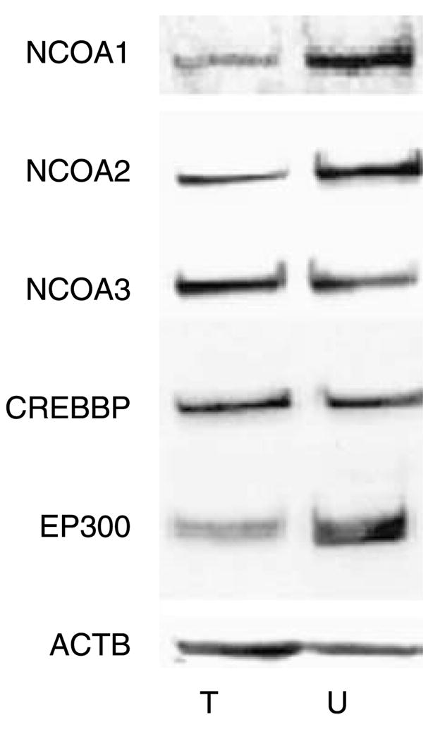
Urothelial carcinoma (UC) of the bladder is approximately three times more common in men than women. While the etiology for this gender difference in incidence remains unknown, a role for androgen receptor (AR) signaling has been suggested. The mechanisms by which AR activity is regulated in UC cells, however, are largely elusive. Here, we explore the significance of coregulators that are critical for the formation of a functional AR transcriptional complex, in UC cells. Using two AR-positive UC cell lines, TCC-SUP and UMUC3, we demonstrate the expression of the coactivators NCOA1, NCOA2, NCOA3, CREBBP, and EP300 in UC cells. small interfering RNA-mediated knockdown of the AR or any of these coactivators markedly impacted cell viability and abrogated androgen-dependent cell proliferation. Noteworthy, contrary to AR-positive prostate cancer cells, expression of these AR-associated coactivators was not androgen regulated in UC cells. To assess the clinical relevance of coactivator expression, we performed immunohistochemistry on paraffin-embedded sections from 55 patients with UC of the bladder. We found that while 24 out of 55 (44%) of tumors expressed the AR, each of the coactivators was expressed by 85-100% of the bladder cancers. Moreover, we noted a significant downregulation of NCOA1 expression in tumors versus adjacent, non-tumor bladder urothelium, with a mean of 68% (range 0-100) of tumor cells demonstrating NCOA1 staining versus a mean of 81% (range 0-90) of non-tumor cells (P=0.03). Taken together, our data suggest an important role for AR-associated coactivators in UC and point toward differences in the regulation of AR activity between bladder and prostate cancer cells.


Conflict of interest statement

Declaration of interest

The authors declare that there is no conflict of interest that would prejudice the impartiality of this scientific work.

Figures

Figure 1
Figure 1
AR expression in UC cell lines. (A) Cell extracts from TCC-SUP (T) and UMUC3 (U) cells were subjected to immunoblotting using an antibody directed against the AR. (B) Cells were cultured in the presence or absence of the synthetic androgen R1881 (1 nM) for 48 h. Western blot analysis was performed to assess AR expression. To evaluate potential intersample loading differences, blots were stripped and reprobed with an antibody recognizing β-actin (ACTB). Blots representative of three separate experiments are shown.
Figure 2
Figure 2
Androgen-stimulated cell viability in AR-positive UC cell lines. Cells were seeded in medium supplemented with CSS. Forty-eight hours later, medium was changed and cells were treated with 1 nM R1881 (black columns) or ethanol vehicle (gray columns). Forty-eight and 96 h later, cell viability was assessed by means of an MTS assay reading absorbance at 490 nm. Columns and vertical bars represent the mean ± S.E.M. of five individual measurements. *Statistically significant difference compared with untreated cells, P<0.05.
Figure 3
Figure 3
Expression of AR core coactivators in AR-positive UC cell lines. Whole-cell lysates from TCC-SUP (T) and UMUC3 (U) cells grown in their regular culture medium were subjected to western blotting using antibodies directed against NCOA1, NCOA2, NCOA3, CREBBP, and EP300. To assess potential intersample loading differences, blots were stripped and reprobed with an antibody recognizing β-actin (ACTB). Blots representative of three separate experiments are shown.
Figure 4
Figure 4
Importance of AR coactivator expression for UC cell viability. Cells were transfected with siRNA against the AR or AR coactivators (black columns) or with control siRNAs (gray columns). Twelve to 16 h after transfection, medium was changed. Forty-eight and 96 h later, cell viability was assessed by means of an MTS assay, reading absorbance at 490 nm. Columns and vertical bars represent the mean±S.E.M. of five individual measurements. *Statistically significant difference compared with control, P<0.05.
Figure 5
Figure 5
Compensatory effect of siRNA-mediated knockdown of AR coactivators on expression of functionally related coactivators in UC cells. TCC-SUP and UMUC3 cells were transfected with siRNA targeting coactivator expression or with control siRNA (c). On the next day, medium was replaced. Ninety-six hours later, cell lysates were prepared and subjected to immunoblot analysis using antibodies targeted against NCOA1, NCOA2, NCOA3, CREBBP, EP300, and β-actin.
Figure 6
Figure 6
Effect of siRNA transfection on coactivator protein expression in UC cells. TCC-SUP and UMUC3 cells were transfected with siRNA targeting AR or AR coactivator expression or with control siRNA (c). On the next day, medium was replaced. Ninety-six hours later, cell lysates were prepared and blotted for the AR, NCOA1, NCOA2, NCOA3, CREBBP, EP300, and β-actin (ACTB).
Figure 7
Figure 7
Loss of AR or AR coactivator expression in UC cells does not induce apoptosis. Cell lysates were obtained from (A) TCC-SUPP and (B) UMUC3 cells transfected with siRNAs targeting the AR, AR coactivators, or control siRNAs (c), as described. Western blotting for expression of the cleavage fragment of PARP was performed. Cell lysates obtained from TCC-SUP and UMUC3 cells treated with 0, 1, or 20 ng/ml staurosporine (Stauro) were similarly evaluated (right). To assess potential intersample loading differences, blots were stripped and reprobed with an antibody recognizing β-actin.
Figure 8
Figure 8
AR coactivators are important for androgen-dependent proliferation of UMUC3 cells. UMUC3 cells seeded in medium supplemented with CSS were transfected with siRNAs targeting the AR, AR coactivators, or non-targeting control siRNAs (c). One day after transfection, medium was changed and cells were treated with 1 nM R1881 (black columns) or ethanol vehicle (gray columns). Ninety-six hours later, cell viability was assessed by means of an MTS assay, reading absorbance at 490 nm. Columns and vertical bars represent the mean±S.E.M. of five individual measurements. *Statistically significant difference compared with control, P<0.05.
Figure 9
Figure 9
AR coactivator expression is not affected by androgen stimulation in UC cells. (A) TCC-SUP and UMUC cells were seeded in medium supplemented with CSS. Two days later, medium was changed and cells were treated with 1 nM R1881 or ethanol vehicle for 48 h. Total protein extracts were prepared, and equal amounts of protein were analyzed by western blotting using antibodies directed against NCOA1, NCOA2, NCOA3, CREBBP, and EP300. To assess potential intersample loading differences, blots were stripped and reprobed with an antibody recognizing β-actin. Blots representative of three separate experiments are shown. (B) TCC-SUP and UMUC cells were seeded in medium supplemented with CSS. Two days later, medium was changed and cells were treated with 1 nM R1881 (black bars) or ethanol vehicle (gray bars) for 48 h. RNA was isolated and converted into cDNA. Real-time PCR was performed using primers specific for NCOA1, NCOA2, NCOA3, CREBBP, and EP300. mRNA levels were normalized with the values obtained from GAPDH expression. mRNA levels are expressed as relative expression values, taking the value obtained from one batch of cells treated with vehicle as 1. Columns, mean values obtained from biological triplicates; bars, S.E.M. (C) AR coactivator expression is independent of AR expression in UC cells. TCC-SUP and UMUC3 cells were transfected with siRNA targeting the AR or with control siRNA (c). On the next day, medium was replaced. Ninety-six hours later, cell lysates were prepared and blotted for the AR, NCOA1, NCOA2, NCOA3, CREBBP, EP300, and β-actin (ACTB).
Figure 10
Figure 10
Expression of AR coactivators and the AR in human UC tumors. Serial tissue sections from a patient with stage pT3 UC of the bladder are shown. (A) H&E section demonstrating areas of invasive tumor (asterisk), carcinoma in situ (thin arrow), and non-tumor urothelium (thick arrow). Scale bar, 50 μm. Results of immunohistochemical staining for (B) NCOA1, (C) NCOA2, (D) NCOA3, (E) CREBBP, (F) EP300, and (G) AR. All images represent original magnification ×200.

Similar articles

Cited by

References

    1. Aarnisalo P, Palvimo JJ, Jänne OA. CREB-binding protein in androgen receptor-mediated signaling. PNAS. 1998;95:2122–2127. - PMC - PubMed
    1. Agoulnik IU, Vaid A, Bingman WE, III, Erdeme H, Frolov A, Smith CL, Ayala G, Ittmann MM, Weigel NL. Role of SRC-1 in the promotion of prostate cancer cell growth and tumor progression. Cancer Research. 2005;65:7959–7967. - PubMed
    1. Agoulnik IU, Vaid A, Nakka M, Alvarado M, Bingman WE, III, Erdem H, Frolov A, Smith CL, Ayala GE, Ittmann MM, et al. Androgens modulate expression of transcription intermediary factor 2, an androgen receptor coactivator whose expression level correlates with early biochemical recurrence in prostate cancer. Cancer Research. 2006;66:10594–10602. - PubMed
    1. Boorjian S, Ugras S, Mongan NP, Gudas LJ, You X, Tickoo SK, Scherr DS. Androgen receptor expression is inversely correlated with pathologic tumor stage in bladder cancer. Urology. 2004;64:383–388. - PubMed
    1. Boorman GA. Animal model of human disease; carcinoma of the ureter and urinary bladder. American Journal of Pathology. 1977;88:251–254. - PMC - PubMed

Publication types

MeSH terms