. 2008 Nov 21;103(11):1289-99.
doi: 10.1161/01.RES.0000338496.95579.56. Epub 2008 Oct 9.

Stim1 and Orai1 mediate CRAC currents and store-operated calcium entry important for endothelial cell proliferation

Affiliations

Stim1 and Orai1 mediate CRAC currents and store-operated calcium entry important for endothelial cell proliferation

Iskandar F Abdullaev et al. Circ Res. .

Abstract

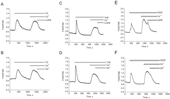
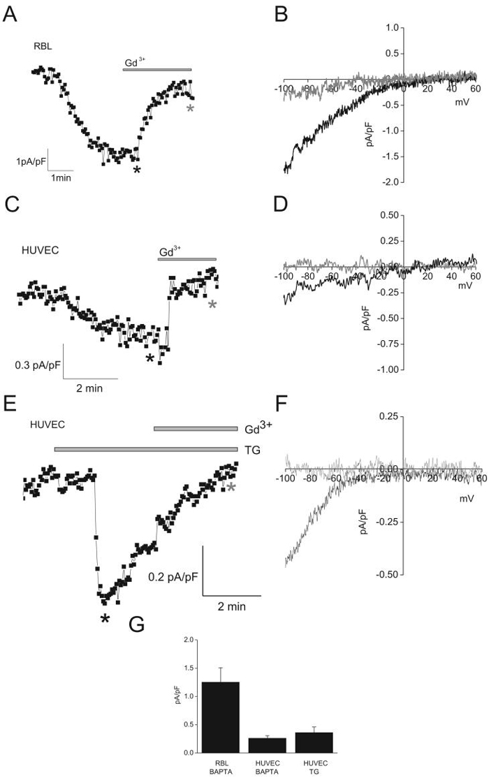
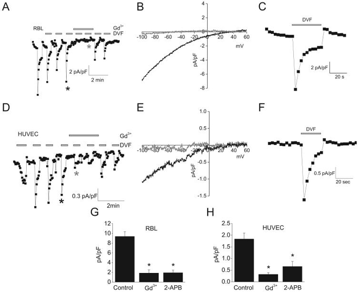
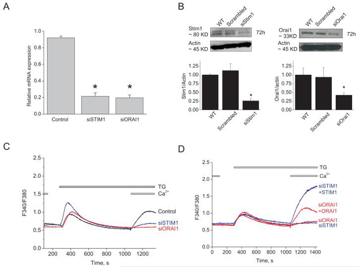
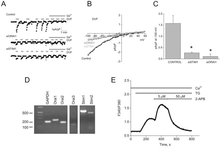
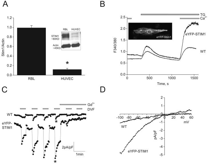
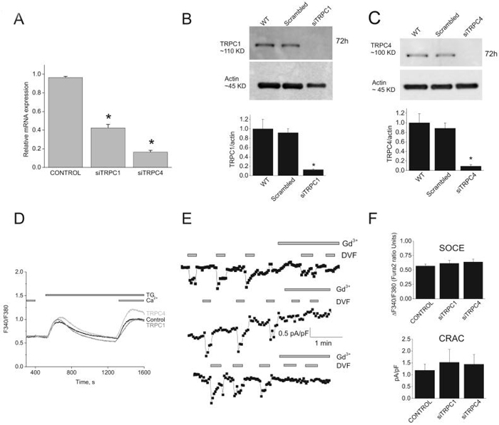
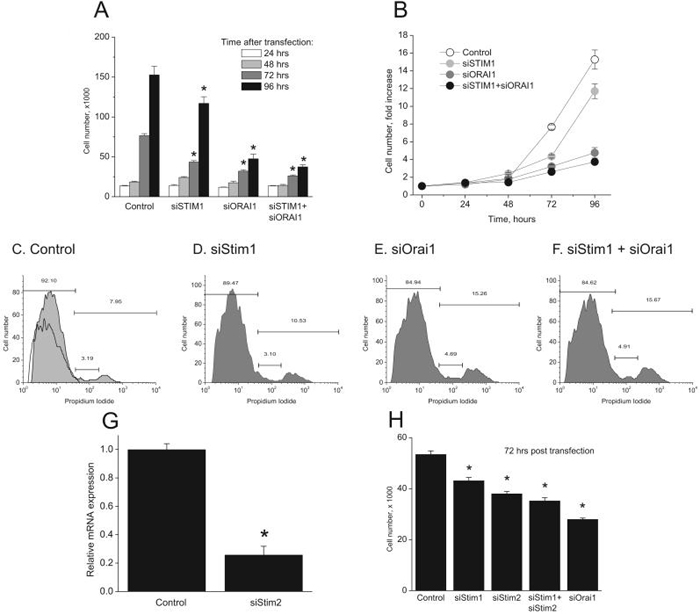
Recent breakthroughs in the store-operated calcium (Ca(2+)) entry (SOCE) pathway have identified Stim1 as the endoplasmic reticulum Ca(2+) sensor and Orai1 as the pore forming subunit of the highly Ca(2+)-selective CRAC channel expressed in hematopoietic cells. Previous studies, however, have suggested that endothelial cell (EC) SOCE is mediated by the nonselective canonical transient receptor potential channel (TRPC) family, TRPC1 or TRPC4. Here, we show that passive store depletion by thapsigargin or receptor activation by either thrombin or the vascular endothelial growth factor activates the same pathway in primary ECs with classical SOCE pharmacological features. ECs possess the archetypical Ca(2+) release-activated Ca(2+) current (I(CRAC)), albeit of a very small amplitude. Using a maneuver that amplifies currents in divalent-free bath solutions, we show that EC CRAC has similar characteristics to that recorded from rat basophilic leukemia cells, namely a similar time course of activation, sensitivity to 2-aminoethoxydiphenyl borate, and low concentrations of lanthanides, and large Na(+) currents displaying the typical depotentiation. RNA silencing of either Stim1 or Orai1 essentially abolished SOCE and I(CRAC) in ECs, which were rescued by ectopic expression of either Stim1 or Orai1, respectively. Surprisingly, knockdown of either TRPC1 or TRPC4 proteins had no effect on SOCE and I(CRAC). Ectopic expression of Stim1 in ECs increased their I(CRAC) to a size comparable to that in rat basophilic leukemia cells. Knockdown of Stim1, Stim2, or Orai1 inhibited EC proliferation and caused cell cycle arrest at S and G2/M phase, although Orai1 knockdown was more efficient than that of Stim proteins. These results are first to our knowledge to establish the requirement of Stim1/Orai1 in the endothelial SOCE pathway.


Figures

Figure 1
Figure 1. Thapsigargin activates SOCE in HUVECs
A and B, store depletion by 2μmol/L thapsigargin (TG) induces SOCE that is inhibited by 30μmol/L 2-APB (A)and 10 μmol/L Gd3+ (B).C and D, thrombin (100nmol/L) induces SOCE and is inhibited by similar concentrations of 2-APB (C) and Gd3+ (D).E and F, Similar results were obtained with VEGF (100ng/mL). Data in each panel is an average of 4-12 cells and representative of at least 3 independent experiments.
Figure 2
Figure 2. ICRAC in HUVEC and RBL cells
A and C, ICRAC activation upon intracellular dialysis with BAPTA in RBL and HUVEC, respectively. E, ICRAC activation upon extracellular application of thapsigargin (TG; 2μmol/L). Ramps from -100mV to +60mV were taken every 3sec, and data points from each ramp were taken at -100mV and plotted. B, D and F, representative ramps taken at time points marked by asterisks in A, C and E, respectively. Black asterisks, before application of Gd3+; grey asterisks, after application of 10μmol/L Gd3+. G, Statistical analysis of maximal current values at -100mV.
Figure 3
Figure 3. ICRAC in DVF conditions
A and D, Na+ currents recorded in response to repetitive pulses (~ every min) of DVF bath solutions in RBL and HUVEC, respectively. After whole cell mode, ramps from -100mV to +60mV were applied, and data points taken at -100mV and plotted. 10μmol/L Gd3+ was added where indicated. B and E, representative ramps of DVF-induced Na+ currents in RBL (B) and HUVEC (E). Ramps shown were taken at time points indicated by asterisks in A and D, respectively. C and F, close-up of DVF-induced Na+ currents (black asterisks in A and D) in RBL and HUVEC. G and H, mean values of maximal Na+ currents at -100 mV before, after application of 10 μmol/L Gd3+ and 30 μmol/L 2-APB in RBL (G) and HUVEC (H).
Figure 4
Figure 4. SOCE in HUVECs is mediated by Stim1 and Orai1
A, quantitative PCR data showing decreased expression of Orai1 and Stim1 mRNA in silenced cells compared to control cells (scrambled siRNA), 72 hrs post-transfection (n=3; one way ANOVA, *p<0.05). B, Western blots and densitometry showing significant down-regulation of Stim1 (left) and Orai1 (right) proteins 72 hrs after siRNA transfection. C, Stim1 and Orai1 knockdown inhibits SOCE in response to thapsigargin. D, SOCE in Stim1- and Orai1-silenced cells can be rescued by overexpression of eYFP-Stim1 and CFP-Orai1, respectively. All traces shown represent average data from 7-25 cells from at least 3 independent experiments.
Figure 5
Figure 5. Stim1 and Orai1 mediate ICRAC in HUVECs
A, siRNA against either Stim1 or Orai1 in HUVECs greatly reduced ICRAC recorded under DVF conditions compared to control scrambled siRNA-transfected cells. B, representative ramps taken at time points indicated by asterisks in A. C, Summary of the whole-cell data recorded from control-, Stim1- and Orai1-siRNA transfected HUVECs. D, mRNA expression of Stim and Orai isoforms in HUVECs. E, Ca2+ imaging showing thapsigargin-activated SOCE, its potentiation with 5μmol/L 2-APB and subsequent inhibition by 50μmol/L 2-APB; trace is average of 22 cells and representative of 4 independent experiments. (one way ANOVA, *p<0.05).
Figure 6
Figure 6. Small ICRAC in HUVECs are due to low expression levels of Stim1
A, western blots showing low Stim1 protein levels in HUVECs compared with RBL cells. Ectopic expression of 1μg of eYFP-Stim1 cDNA plasmid (B; see inset) in HUVECs dramatically increased SOCE (B) and ICRAC (C). D, Representative ramps of ICRAC in wild type (WT) and eYFP-Stim1-expressing HUVECs were taken from C, where indicated by asterisks.
Figure. 7
Figure. 7. TRPC1 and TRPC4 knockdown has no effect on SOCE and ICRAC in HUVECs
A, quantitative PCR showing decreased expression of TRPC1 and TRPC4 mRNA in silenced cells compared to control cells (scrambled siRNA), 72 hrs after transfection. B and C, western blots and statistical analysis of densitometry on TRPC1 and TRPC4 protein knockdown. D, TRPC1 and TRPC4 silencing has no effect on SOCE in response to thapsigargin in HUVECs. Data are representative of 12 independent experiments. E, ICRAC recorded in DVF conditions from control-, TRPC1- and TRPC4-siRNA transfected HUVECs. F, statistical analysis of Ca2+ entry measured by Fura2 (top) and ICRAC (bottom) from control-, TRPC1- and TRPC4-siRNA transfected HUVECs. (One way ANOVA, *p<0.05).
Figure. 8
Figure. 8. Stim1, Stim2 and Orai1 knockdown inhibit proliferation in HUVECs
A, Four groups of cells were transfected with the following siRNA: control, Stim1, Orai1, Stim1 + Orai1. At time 0, approximately 10,000 cells were seeded per well (9.6 cm2) in triplicate. At times indicated, cells were detached and total number of cells per well counted after trypan blue exclusion. B, Data in “A” are represented as fold-increase of cell number compared to time 0. Cell cycle analysis was performed 3 days post-transfection with siRNA using Propidium Iodide (PI) staining with flow cytometry in control- (C), Stim1- (D), Orai1- (E), and Stim1 + Orai1- (F) siRNA transfected HUVECs. Non PI stained control in shown in panel C. G, quantitative PCR showing Stim2 mRNA knockdown, 72 hours after transfection with specific siRNA. H, EC proliferation at 72hours post-transfection with indicated siRNA using the same protocol described in A. Data are representative of four independent experiments (one way ANOVA, *p<0.05).

Comment in

References

    1. Putney JW., Jr. A model for receptor-regulated calcium entry. Cell Calcium. 1986 Feb;7(1):1–12. - PubMed
    1. Smyth JT, Dehaven WI, Jones BF, Mercer JC, Trebak M, Vazquez G, Putney JW., Jr. Emerging perspectives in store-operated Ca(2+) entry: Roles of Orai, Stim and TRP. Biochim Biophys Acta. 2006 Sep 5; - PubMed
    1. Hoth M, Penner R. Depletion of intracellular calcium stores activates a calcium current in mast cells. Nature. 1992 Jan 23;355(6358):353–356. - PubMed
    1. Parekh AB, Putney JW., Jr. Store-operated calcium channels. Physiol Rev. 2005 Apr;85(2):757–810. - PubMed
    1. Feske S, Gwack Y, Prakriya M, Srikanth S, Puppel SH, Tanasa B, Hogan PG, Lewis RS, Daly M, Rao A. A mutation in Orai1 causes immune deficiency by abrogating CRAC channel function. Nature. 2006 May 11;441(7090):179–185. - PubMed

Publication types

MeSH terms