. 2007;2(4):451-469.
doi: 10.1586/17469872.2.4.451.

Mechanism of UV-related carcinogenesis and its contribution to nevi/melanoma

Affiliations

Mechanism of UV-related carcinogenesis and its contribution to nevi/melanoma

Brozyna Anna et al. Expert Rev Dermatol. 2007.

Abstract

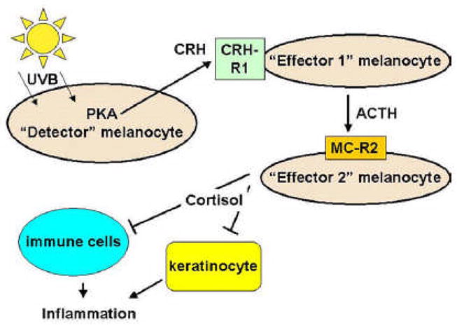
Melanoma consists 4-5 % of all skin cancers, but it contributes to 71-80 % of skin cancers deaths. UV light affects cell and tissue homeostasis due to its damaging effects on DNA integrity and modification of expression of a plethora of genes. DNA repair systems protect cells from UV-induced lesions. Several animal models of melanoma have been developed (Xiphophorus, Opossum Monodelphis domestica, mouse models and human skin engrafts into other animals). This review discusses possible links between UV and genes significantly related to melanoma but does not discuss melanoma genetics. These include oncogenes, tumor suppressor genes, genes related to melanocyte-keratinocyte and melanocyte-matrix interaction, growth factors and their receptors, CRH, ACTH, α-MSH, glucocorticoids, ID1, NF-kappaB and vitamin D3.


Figures

FIGURE 1
FIGURE 1
Three major setting of UV induced melanoma: A) intermittent sun exposure in nevus susceptible patient; B) multiple sun burns during childhood-aldolescence; and C) Solar damaged skin with multiple lentigines.
FIGURE 2
FIGURE 2. Melanocytic hyperplasia occurring in sun damaged skin
Formation of solar lentigines, (hyperpigmented, club shaped rete ridges overlying abundant solar elastotic material) after chronic sun exposure (A). Melanocytes are increased in density in sun-damaged skin (B) as well as within solar lentigines (C). Mart-1 labeling (A and C) demonstrates that both regions have an increase in melanocytes density as well as enlargement of many of the melanocytes.
FIGURE 3
FIGURE 3. Lentigo maligna melanoma arising on sun damaged skin
A–B. A typical lentigo maligna melanoma (LMM- invasive melanoma, dark central region) arising in long standing patch of lentigo maligna (melanoma in situ, large irregular brown patch) occurring in sun damaged skin of an elderly female. C. Early vertical growth phase LMM characterized by small nest of melanoma within the papillary dermis found in association with confluent nests of LM found along the dermal-epidermal junction. D. Antibodies to CD117/c-kit demonstrate strong and intense expression of the both in situ and invasive components.
FIGURE 4
FIGURE 4. Melanoma arising in association with melanocytic nevus
A. Superificial spreading melanoma arising in the background of scattered small common melanocytic nevus cells. B. Scanning magnification shows vertical growth phase melanoma (left side) arising in association with intradermal congential pattern melanocytic nevus (right side). C. Melanoma in situ (large junctional nests of atypical melanocytes) overlying cytologically banal dermal melanocytes (arrow) associated with lymphocytic inflammatory host response.
FIGURE 5
FIGURE 5
p53 mutant keratinocyte clones and mutant melanocytes occurring in sun damaged skin. Illustrated herein are mutant keratinocytes of solar lentigo (A & C) and putative p53 mutant melanocytes of melanoma in situ (B & D) arising in sun damaged skin.
FIGURE 6
FIGURE 6
Hypothetical model of inter-melanocyte interactions through local HPA axis mediators. UVB stimulates production of CRH in “detector” melanocyte [121]. CRH stimulates “effector 1” melanocyte to produce ACTH. In turn, ACTH in stimulates steroid production by “effector 2” melanocyte [124]. Acting together, ACTH and steroids suppress local inflammatory reaction proceeding with the participation of keratinocytes [149] and immune cells [150].

References

    1. Slominski A, Pawelek J. Animals under the sun: Effects of UV radiation on mammalian skin. Clin Dermatol. 1998;16:503–515. - PubMed
    1. Brenner M, Degitz K, Besch R, Berking C. Differential expression of melanoma-associated growth factors in keratinocytes and fibroblasts by ultraviolet A and ultraviolet B radiation. Br J Dermatol. 2005;153(4):733–9. “considerable interest” Extremely convincing experiments showing that synthesis of growth factors for melanocytes is dependent on cell type and UV wavelength. - PubMed
    1. Norval M. The mechanisms and consequences of ultraviolet-induced immunosuppression. Prog Biophys Mol Biol. 2006;92(1):108–18. - PubMed
    1. Jhappan C, Noonan FP, Merlino G. Ultraviolet radiation and cutaneous malignant melanoma. Oncogene. 2003;22(20):3099–112. - PubMed
    1. Mariutti G, Matzeu M. Measurement of ultraviolet radiation emitted from welding arcs. Health Phys. 1988;54(5):529–32. - PubMed

LinkOut - more resources