Instrumented posterior lumbar interbody fusion in adult spondylolisthesis
- PMID: 18846411
- PMCID: PMC2628248
- DOI: 10.1007/s11999-008-0511-1
Instrumented posterior lumbar interbody fusion in adult spondylolisthesis
Abstract
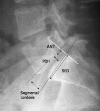
It is unclear whether using artificial cages increases fusion rates compared with use of bone chips alone in posterior lumbar interbody fusion for patients with lumbar spondylolisthesis. We hypothesized artificial cages for posterior lumbar interbody fusion would provide better clinical and radiographic outcomes than bone chips alone. We assumed solid fusion would provide good clinical outcomes. We clinically and radiographically followed 34 patients with spondylolisthesis having posterior lumbar interbody fusion with mixed autogenous and allogeneic bone chips alone and 42 patients having posterior lumbar interbody fusion with implantation of artificial cages packed with morselized bone graft. Patients with the artificial cage had better functional improvement in the Oswestry disability index than those with bone chips alone, whereas pain score, patient satisfaction, and fusion rate were similar in the two groups. Postoperative disc height ratio, slip ratio, and segmental lordosis all decreased at final followup in the patients with bone chips alone but remained unchanged in the artificial cage group. The functional outcome correlated with radiographic fusion status. We conclude artificial cages provide better functional outcomes and radiographic improvement than bone chips alone in posterior lumbar interbody fusion for lumbar spondylolisthesis, although both techniques achieved comparable fusion rates.
Level of evidence: Level III, therapeutic study. See the Guidelines for Authors for a complete description of levels of evidence.
Figures


Similar articles
-
Microscopic anterior foraminal decompression combined with anterior lumbar interbody fusion.Spine J. 2013 Oct;13(10):1190-9. doi: 10.1016/j.spinee.2013.07.458. Epub 2013 Oct 2. Spine J. 2013. PMID: 24094988
-
Posterior lumbar interbody fusion for degenerative spondylolisthesis: restoration of sagittal balance using insert-and-rotate interbody spacers.Spine J. 2005 Mar-Apr;5(2):170-9. doi: 10.1016/j.spinee.2004.05.257. Spine J. 2005. PMID: 15749617 Clinical Trial.
-
Outcomes of autograft alone versus PEEK+ autograft interbody fusion in the treatment of adult lumbar isthmic spondylolisthesis.Clin Neurol Neurosurg. 2017 Apr;155:1-6. doi: 10.1016/j.clineuro.2017.01.020. Epub 2017 Jan 31. Clin Neurol Neurosurg. 2017. PMID: 28187368
-
[Lumbar interbody fusion. Biomechanical significance for the spine].Neurol Neurochir Pol. 1998 Sep-Oct;32(5):1247-59. Neurol Neurochir Pol. 1998. PMID: 10463237 Review. Polish.
-
Intervertebral cages for degenerative spinal diseases.Spine J. 2003 Jul-Aug;3(4):301-9. doi: 10.1016/s1529-9430(03)00004-4. Spine J. 2003. PMID: 14589191 Review.
Cited by
-
Surgical options for lumbar spinal stenosis.Cochrane Database Syst Rev. 2016 Nov 1;11(11):CD012421. doi: 10.1002/14651858.CD012421. Cochrane Database Syst Rev. 2016. PMID: 27801521 Free PMC article.
-
Comparing Lumbar Spine Cages and Bone Grafts in Spinal Arthrodesis: A Meta-Analysis of Clinical Outcomes.Cureus. 2025 Jan 6;17(1):e77017. doi: 10.7759/cureus.77017. eCollection 2025 Jan. Cureus. 2025. PMID: 39912040 Free PMC article.
-
Comparison of the PEEK cage and an autologous cage made from the lumbar spinous process and laminae in posterior lumbar interbody fusion.BMC Musculoskelet Disord. 2016 Aug 30;17(1):374. doi: 10.1186/s12891-016-1237-y. BMC Musculoskelet Disord. 2016. PMID: 27577978 Free PMC article. Clinical Trial.
-
Biomechanical comparison of polyetheretherketone rods and titanium alloy rods in transforaminal lumbar interbody fusion: a finite element analysis.BMC Surg. 2024 May 29;24(1):169. doi: 10.1186/s12893-024-02462-8. BMC Surg. 2024. PMID: 38811965 Free PMC article.
-
Pedicle-Screw-Based Dynamic Systems and Degenerative Lumbar Diseases: Biomechanical and Clinical Experiences of Dynamic Fusion with Isobar TTL.ISRN Orthop. 2013 Jan 21;2013:183702. doi: 10.1155/2013/183702. eCollection 2013. ISRN Orthop. 2013. PMID: 25031874 Free PMC article.
References
-
- {'text': '', 'ref_index': 1, 'ids': [{'type': 'PubMed', 'value': '10505503', 'is_inner': True, 'url': 'https://pubmed.ncbi.nlm.nih.gov/10505503/'}]}
- Agazzi S, Reverdin A, May D. Posterior lumbar interbody fusion with cages: an independent review of 71 cases. J Neurosurg. 1999;91:186–192. - PubMed
-
- {'text': '', 'ref_index': 1, 'ids': [{'type': 'DOI', 'value': '10.1097/00007632-200307150-00016', 'is_inner': False, 'url': 'https://doi.org/10.1097/00007632-200307150-00016'}, {'type': 'PubMed', 'value': '12865845', 'is_inner': True, 'url': 'https://pubmed.ncbi.nlm.nih.gov/12865845/'}]}
- Akamaru T, Kawahara N, Tim Yoon S, Minamide A, Su Kim K, Tomita K, Hutton WC. Adjacent segment motion after a simulated lumbar fusion in different sagittal alignments: a biomechanical analysis. Spine. 2003;28:1560–1566. - PubMed
-
- {'text': '', 'ref_index': 1, 'ids': [{'type': 'PubMed', 'value': '12401914', 'is_inner': True, 'url': 'https://pubmed.ncbi.nlm.nih.gov/12401914/'}]}
- Arai Y, Takahashi M, Kurosawa H, Shitoto K. Comparative study of iliac bone graft and carbon cage with local bone graft in posterior lumbar interbody fusion. J Orthop Surg (Hong Kong). 2002;10:1–7. - PubMed
-
- {'text': '', 'ref_index': 1, 'ids': [{'type': 'PubMed', 'value': '8362324', 'is_inner': True, 'url': 'https://pubmed.ncbi.nlm.nih.gov/8362324/'}]}
- Blumenthal SL, Gill K. Can lumbar spine radiographs accurately determine fusion in postoperative patients? Correlation of routine radiographs with a second surgical look at lumbar fusions. Spine. 1993;18:1186–1189. - PubMed
-
- {'text': '', 'ref_index': 1, 'ids': [{'type': 'DOI', 'value': '10.1016/S1529-9430(02)00536-3', 'is_inner': False, 'url': 'https://doi.org/10.1016/s1529-9430(02)00536-3'}, {'type': 'PubMed', 'value': '14589199', 'is_inner': True, 'url': 'https://pubmed.ncbi.nlm.nih.gov/14589199/'}]}
- Brantigan JW, Neidre A. Achievement of normal sagittal plane alignment using a wedged carbon fiber reinforced polymer fusion cage in treatment of spondylolisthesis. Spine J. 2003;3:186–196. - PubMed
Publication types
MeSH terms
LinkOut - more resources
Full Text Sources
Research Materials

