Comparative Study
. 2008 Jun;83(3-4):179-89.
doi: 10.1080/10520290802451085.

Immunohistochemistry in the evaluation of neovascularization in tumor xenografts

Affiliations
Comparative Study

Immunohistochemistry in the evaluation of neovascularization in tumor xenografts

D Wang et al. Biotech Histochem. 2008 Jun.

Abstract

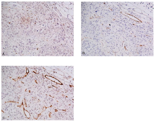
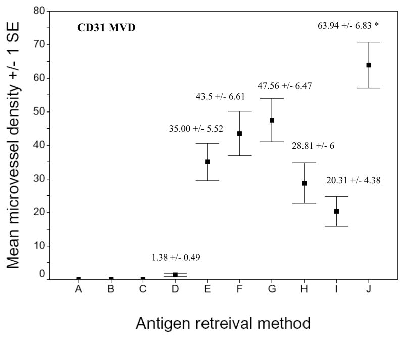
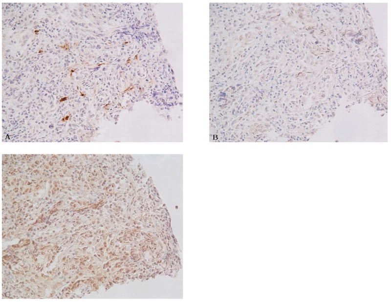
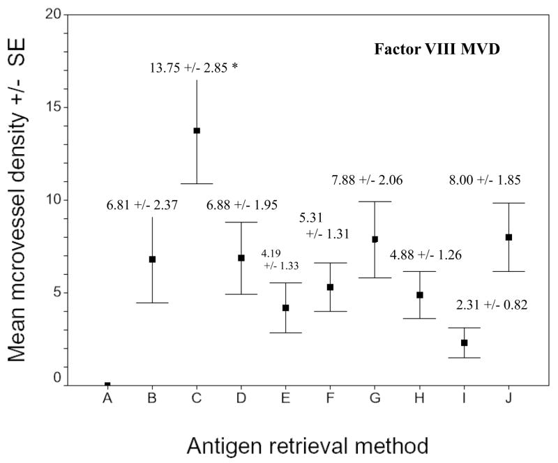
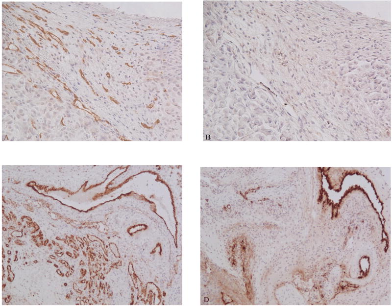
Angiogenesis, or neovascularization, is known to play an important role in the neoplastic progression leading to metastasis. CD31 or Factor VIII-related antigen (F VIII RAg) immunohistochemistry is widely used in experimental studies for quantifying tumor neovascularization in immunocompromised animal models implanted with transformed human cell lines. Quantification, however, can be affected by variations in the methodology used to measure vascularization including antibody selection, antigen retrieval (AR) pretreatment, and evaluation techniques. To examine this further, we investigated the microvessel density (MVD) and the intensity of microvascular staining among five different human tumor xenografts and a mouse syngeneic tumor using anti-CD31 and F VIII RAg immunohistochemical staining. Different AR methods also were evaluated. Maximal retrieval of CD31 was achieved using 0.5 M Tris (pH 10) buffer, while maximum retrieval of F VIII RAg was achieved using 0.05% pepsin treatment of tissue sections. For each optimized retrieval condition, anti-CD31 highlighted small vessels better than F VIII RAg. Furthermore, the MVD of CD31 was significantly greater than that of F VIII RAg decorated vessels (p<0.001). The choice of antibody and AR method has a significant affect on immunohistochemical findings when studying angiogenesis. One also must use caution when comparing studies in the literature that use different techniques and reagents.


Figures

Fig. 1
Fig. 1
Comparision of CD31 immunohistochemistry using various AR methods on serial sections of the squamous carcinoma xenograft. A) 0.05% pepsin treated section. B) 0.01 M Citric acid (pH 6) treated section. C) 0.5 M Tris buffer (pH 10) treated section. All panels 200 X.
Fig. 2
Fig. 2
Microvessel density of CD31 stained sections of xenografts using different AR methods. A) No treatment. B) 0.1% trypsin. C) 0.05% pepsin. D) 0.01 M glycine (pH 3). E) 0.01 M sodium citric buffer (pH 6). F) 0.05 M borate buffer (pH 8). G) 0.01 M Tris-EDTA buffer (pH 9). H) AR10 solution (pH 10). I) 0.01 M Tris/0.05% Tween 20 (pH 10). J) 0.5 M Tris (pH 10). Asterisk indicates that the CD31 microvellel development seen with 0.5 M Tris (pH 10) was significantly greater than that obtained using the other AR methods (p < 0.01).
Fig. 3
Fig. 3
Comparision of Factor VIII IHC by various AR methods on serial sections of colon adenocacinoma xenograft. A) 0.05% pepsin treated section. B) 0.01 M vitric acid (pH 6) treated section. C) 0.5 M Tris buffer (pH 10) treated section. All figures 200 X.
Fig. 4
Fig. 4
Microvessel density of F VIII RAg stained sections of xenografts using different AR methods. A) No treatment. B) 0.1% trypsin. C = 0.05% pepsin. D) 0.01M glycine (pH 3). E) 0.01 M sodium citric buffer (pH 6). F) 0.05 M borate buffer (pH 8). G) 0.01 M Tris-EDTA buffer (pH 9). H) AR10 solution (pH 10). I) 0.01 M Tris/0.05% Tween 20 (pH 10). J) 0.5 M Tris (pH 10). Asterisk indicates that the F VIII RAg microvessel development seen after pepsin digestion was significantly greater than that obtained by using the other AR methods (p < 0.01).
Fig. 5
Fig. 5
Comparision of anti-CD31 and anti-F VIII RAg stains using the optimized AR method. Serial sections from MDA-MB-231 breast cancer xenograft (A and B) and syngeneic breast cancer (C and D). Sections were treated with 0.5 M Tris buffer (pH 10) followed by the CD31 immunohistochemistry (A and C). Sections were treated with 0.05% pepsin followed by F VIII RAg immunohistochemistry (B and D). Anti-CD31 produced better microvessel staining compared to anti-F VIII RAg staining. A and B: 200 X; C and D 100 X.

References

    1. Behrem S, Zarkovic K, Eskinja N, Jonjic N. Endoglin is a better marker than CD31 in evaluation of angiogenesis in glioblastoma. Croat Med J. 2005;46:417–22. - PubMed
    1. Cattoretti G, Pileri S, Parravicini C, Becker MH, Poggi S, Bifulco C, Key G, D’Amato L, Sabattini E, Feudale E. Antigen unmasking on formalin-fixed, paraffin-embedded tissue sections. J Pathol. 1993;171:83–98. - PubMed
    1. Chi JT, Chang HY, Haraldsen G, Jahnsen FL, Troyanskaya OG, Chang DS, Wang Z, Rockson SG, van de Rijn M, Botstein D, Brown PO. Endothelial cell diversity revealed by global expression profiling. Proc Natl Acad Sci U S A. 2003;100:10623–8. - PMC - PubMed
    1. Folberg R, Hendrix MJ, Maniotis AJ. Vasculogenic mimicry and tumor angiogenesis. Am J Pathol. 2000;156:361–81. - PMC - PubMed
    1. Fox SB. Tumour angiogenesis and prognosis. Histopathology. 1997;30:294–301. - PubMed

Publication types

MeSH terms