. 2008 Nov;48(5):1644-54.
doi: 10.1002/hep.22498.

Translocation of iron from lysosomes into mitochondria is a key event during oxidative stress-induced hepatocellular injury

Affiliations

Translocation of iron from lysosomes into mitochondria is a key event during oxidative stress-induced hepatocellular injury

Akira Uchiyama et al. Hepatology. 2008 Nov.

Abstract

Iron overload exacerbates various liver diseases. In hepatocytes, a portion of non-heme iron is sequestered in lysosomes and endosomes. The precise mechanisms by which lysosomal iron participates in hepatocellular injury remain uncertain. Here, our aim was to determine the role of intracellular movement of chelatable iron in oxidative stress-induced killing to cultured hepatocytes from C3Heb mice and Sprague-Dawley rats. Mitochondrial polarization and chelatable iron were visualized by confocal microscopy of tetramethylrhodamine methylester (TMRM) and quenching of calcein, respectively. Cell viability and hydroperoxide formation (a measure of lipid peroxidation) were measured fluorometrically using propidium iodide and chloromethyl dihydrodichlorofluorescein, respectively. After collapse of lysosomal/endosomal acidic pH gradients with bafilomycin (50 nM), an inhibitor of the vacuolar proton-pumping adenosine triphosphatase, cytosolic calcein fluorescence became quenched. Deferoxamine mesylate and starch-deferoxamine (1 mM) prevented bafilomycin-induced calcein quenching, indicating that bafilomycin induced release of chelatable iron from lysosomes/endosomes. Bafilomycin also quenched calcein fluorescence in mitochondria, which was blocked by 20 microM Ru360, an inhibitor of the mitochondrial calcium uniporter, consistent with mitochondrial iron uptake by the uniporter. Bafilomycin alone was not sufficient to induce mitochondrial depolarization and cell killing, but in the presence of low-dose tert-butylhydroperoxide (25 microM), bafilomycin enhanced hydroperoxide generation, leading to mitochondrial depolarization and subsequent cell death.

Conclusion: Taken together, the results are consistent with the conclusion that bafilomycin induces release of chelatable iron from lysosomes/endosomes, which is taken up by mitochondria. Oxidative stress and chelatable iron thus act as two "hits" synergistically promoting toxic radical formation, mitochondrial dysfunction, and cell death. This pathway of intracellular iron translocation is a potential therapeutic target against oxidative stress-mediated hepatotoxicity.


Figures

Figure 1
Figure 1. Inhibition of bafilomycin-induced intracellular calcein quenching by desferal and starch-desferal in mouse hepatocytes
Mouse hepatocytes were co-loaded with TMRM (100 nM) and calcein-AM (1 μM) and incubated with PI (3 μM), calcein free acid (300 μM) and TMRM (50 nM) in the extracellular medium with and without desferal (1 mM) or starch-desferal (1 mM desferal equivalency), as described in MATERIALS AND METHODS. Green fluorescence of calcein and red fluorescence of TMRM and PI were then imaged by laser scanning confocal microscopy before (0 min) and at 60 and 120 min after no further addition (A, Control), 50 nM bafilomycin (B, Baf), bafilomycin in the presence of desferal (C, Baf+DFO) and bafilomycin in the presence of starch-desferal (D, sBaf+DFO). Note the marked decrease of green calcein fluorescence in the cytosol in B after bafilomycin addition, which did not occur during the control incubation (A) and which was suppressed in the presence of desferal (C) and starch-desferal (D). TMRM fluorescence was maintained under all conditions, and PI did not label nuclei. Each experiment is typical of 3 or more replicates.
Figure 2
Figure 2. Quantitation of calcein quenching after bafilomycin treatment
Mouse hepatocytes were loaded with calcein and treated as described in Fig. 1. Average calcein fluorescence of individual hepatocytes after background subtraction was determined at 60 and 120 min of incubation as the percentage of fluorescence prior to additions (0 min). Baf, bafilomycin; DFO, desferal; sDFO, starch-desferal; *, p < 0.01 compared to other groups (n = 2 to 5 hepatocytes per group).
Figure 3
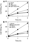
Figure 3. Synergistic cell killing after bafilomycin plus t-BuOOH: protection by desferal and starch-desferal
Viability of mouse hepatocytes was assessed by PI fluorometry, as described in MATERIALS AND METHODS. In A, hepatocytes were exposed to t-BuOOH (25 μM) with and without 60 min of pretreatment with 50 nM bafilomycin (Baf). In B, hepatocytes were treated with desferal (1 mM) or starch-desferal (1 mM desferal equivalency) or no addition prior to bafilomycin plus t-BuOOH treatment. In both panels, “None” represents hepatocytes incubated without any additions. Values are means ± SE from 3 or more hepatocyte isolations.
Figure 4
Figure 4. Mitochondrial depolarization and cell death after bafilomycin plus t-BuOOH: protection by desferal and starch-desferal
Mouse hepatocytes were loaded, as described in Fig. 1, and exposed to 25 μM t-BuOOH alone (A), t-BuOOH plus 50 nM bafilomycin (Baf) (B), t-BuOOH plus bafilomycin after pretreatment with desferal (DFO, 1 mM) (C) and t-BuOOH plus bafilomycin after pretreatment with starch-desferal (DFO, 1 mM desferal equivalency) (D). After t-BuOOH alone (A), note that red mitochondrial TMRM fluorescence was retained and green calcein quenching did not occur. When t-BuOOH was combined with bafilomycin, calcein quenching, loss of TMRM and cellular blebbing occurred within 60 min followed by nuclear PI labeling with 2 h (B). Desferal and starch-desferal prevented calcein quenching, loss of TMRM fluorescence and nuclear labeling with PI (C and D). Each experiment is typical of 3 or more replicates.
Figure 5
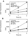
Figure 5. ROS formation after bafilomycin plus t-BuOOH: protection by desferal and starch-desferal
Mouse hepatocytes were incubated with cmH2DCF-DA (10 μM, and fluorescence was measured using a fluorescence plate reader. In A, hepatocytes were exposed to t-BuOOH (25 μM) with and without 60 min pretreatment with 50 nM bafilomycin (Baf) in comparison to bafilomycin alone. In B, hepatocytes were treated with desferal (DFO, 1 mM), starch-desferal (sDFO, 1 mM desferal equivalency) or no addition prior to bafilomycin plus t-BuOOH. In both panels, “Control” represents hepatocytes incubated without any additions. Values are means ± SE from 3 or more hepatocyte isolations.
Figure 6
Figure 6. Calcein quenching in mitochondria and unquenching in lysosomes after bafilomycin
Rat hepatocytes were loaded with 70 kDa rhodamine-dextran (Rhod-Dex) and co-loaded with calcein by cold ester loading/warm incubation, as described in MATERIALS AND METHODS. The hepatocytes were then exposed to no addition (Control) (A), bafilomycin (Baf, 50 nM) (B), bafilomycin after pretreatment with desferal (DFO, 1 mM) (C), and bafilomycin after pretreatment with starch-desferal (sDFO, 1 mM desferal equivalency) (D). Red fluorescence of rhodamine-dextran and green fluorescence of calcein were imaged by laser scanning confocal microscopy. Bafilomycin was added after collection of a baseline image (0 min) and then after 60 and 120 min. Note that mitochondrial calcein fluorescence and lysosomal rhodamine-dextran fluorescence did not change during the control incubation (A). By contrast, mitochondrial calcein was quenched markedly after bafilomycin, whereas lysosomal calcein fluorescence co-localizing with rhodamine-dextran appeared to increase (B). In the presence of desferal (C) and starch-desferal (D), mitochondrial calcein quenching was suppressed, whereas the increase of lysosomal calcein fluorescence appeared to be more marked. Rhodamine-dextran did not leak from lysosomes under any condition. Arrows identify representative lysosomal structures. Each experiment is typical of 3 or more replicates.
Figure 7
Figure 7. Suppression of mitochondrial calcein quenching after bafilomycin by Ru360
Rat hepatocytes were loaded with rhodamine-dextran and calcein, as described in Fig. 6, and exposed to 50 nM bafilomycin (Baf) in the presence of 20 μM Ru360. Note that in comparison to bafilomycin alone (Fig. 6B), mitochondrial calcein quenching was suppressed by Ru360. Arrows identify representative lysosomal structures. One experiment is typical of 3 or more replicates.

Similar articles

Cited by

References

    1. Kehrer JP. The Haber-Weiss reaction and mechanisms of toxicity. Toxicology. 2000 Aug 14;149(1):43–50. - PubMed
    1. Starke PE, Farber JL. Ferric iron and superoxide ions are required for the killing of cultured hepatocytes by hydrogen peroxide. Evidence for the participation of hydroxyl radicals formed by an iron-catalyzed Haber-Weiss reaction. J Biol Chem. 1985 Aug 25;260(18):10099–10104. - PubMed
    1. Kyle ME, Miccadei S, Nakae D, Farber JL. Superoxide dismutase and catalase protect cultured hepatocytes from the cytotoxicity of acetaminophen. Biochem Biophys Res Commun. 1987 Dec 31;149(3):889–896. - PubMed
    1. Dawson TL, Gores GJ, Nieminen AL, Herman B, Lemasters JJ. Mitochondria as a source of reactive oxygen species during reductive stress in rat hepatocytes. Am J Physiol. 1993 Apr;264(4 Pt 1):C961–C967. - PubMed
    1. Nieminen AL, Byrne AM, Herman B, Lemasters JJ. Mitochondrial permeability transition in hepatocytes induced by t-BuOOH: NAD(P)H and reactive oxygen species. Am J Physiol. 1997 Apr;272(4 Pt 1):C1286–C1294. - PubMed

Publication types

MeSH terms