. 2009 Feb;24(2):274-82.
doi: 10.1359/jbmr.081003.

Skeletal cell fate decisions within periosteum and bone marrow during bone regeneration

Affiliations

Skeletal cell fate decisions within periosteum and bone marrow during bone regeneration

Céline Colnot. J Bone Miner Res. 2009 Feb.

Erratum in

  • J Bone Miner Res. 2009 Apr;24(4):758

Abstract

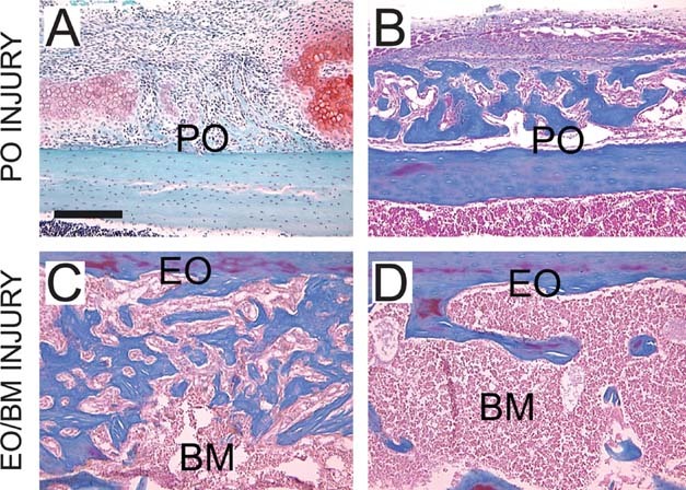
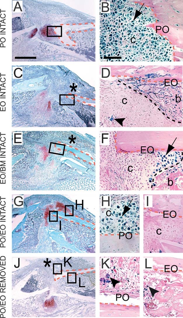
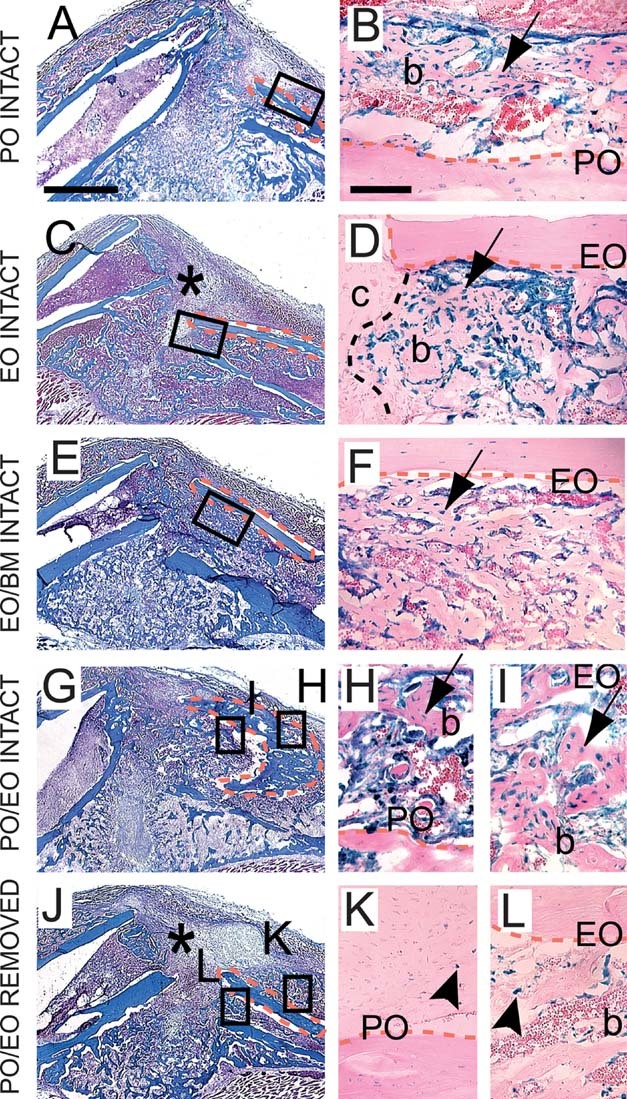
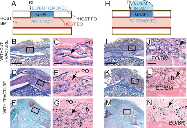
Bone repair requires the mobilization of adult skeletal stem cells/progenitors to allow deposition of cartilage and bone at the injury site. These stem cells/progenitors are believed to come from multiple sources including the bone marrow and the periosteum. The goal of this study was to establish the cellular contributions of bone marrow and periosteum to bone healing in vivo and to assess the effect of the tissue environment on cell differentiation within bone marrow and periosteum. Results show that periosteal injuries heal by endochondral ossification, whereas bone marrow injuries heal by intramembranous ossification, indicating that distinct cellular responses occur within these tissues during repair. [corrected] Next, lineage analyses were used to track the fate of cells derived from periosteum, bone marrow, and endosteum, a subcompartment of the bone marrow. Skeletal progenitor cells were found to be recruited locally and concurrently from periosteum and/or bone marrow/endosteum during bone repair. Periosteum and bone marrow/endosteum both gave rise to osteoblasts, whereas the periosteum was the major source of chondrocytes. Finally, results show that intrinsic and environmental signals modulate cell fate decisions within these tissues. In conclusion, this study sheds light into the origins of skeletal stem cells/progenitors during bone regeneration and indicates that periosteum, endosteum, and bone marrow contain pools of stem cells/progenitors with distinct osteogenic and chondrogenic potentials that vary with the tissue environment.


Figures

FIG. 1
FIG. 1
Periosteal injuries heal by endochondral ossification, whereas endosteal/bone marrow injuries heal by intramembranous ossification. Longitudinal sections through the mouse tibia stained with (A) Safranin-O/Fast Green (SO) and (B-D) trichrome (TC) at (A and C) 7 and (B and D) 14 days after (A and B) periosteal (PO) or (C and D) endosteal/bone marrow (EO/BM) injury. Scale bar = 200 μm.
FIG. 2
FIG. 2
Periosteum and bone marrow/endosteum give rise to osteoblasts/osteocytes during bone graft healing. (Left) Schematic representations of Rosa 26 (blue) or wildtype bone grafts transplanted in the cortex of wildtype host tibias. Longitudinal sections through the mouse tibia stained with TC (middle) and adjacent sections stained with X-gal (right) at 14 days after bone grafting. X-gal-positive osteoblasts and osteocytes (arrow) are found at the intact periosteal surface (A-C), at the intact endosteal surface (D-F), within the marrow cavity (G-I), and at both periosteal and endosteal surfaces in positive controls (J-M), but not in negative controls (N-U). X-gal-positive osteoclasts express β-galactosidase ubiquitously (K, L, T, and U, arrowheads). Dotted orange lines delimit the bone graft. High magnifications correspond to boxed areas. Scale bars: B, E, H, K, O, and S = 1 mm; C, F, I, L, M, P, Q, and T = 100 μm; U = 20 μm.
FIG. 3
FIG. 3
Chondrocytes are primarily derived from the periosteum during healing of nonstabilized fractures. Longitudinal sections through the fracture callus stained with SO (left) and adjacent sections stained with X-gal (right) 10 days after bone grafting and nonstable fracture. (A and B) Cartilage (c) forming at the intact periosteal surface stains positive with X-gal (arrow). Cartilage adjacent to the intact endosteal surface is (C and D) X-gal negative or (E and F) contains few X-gal-positive chondrocytes (arrow) when bone marrow is left intact. (G-I) In positive controls, X-gal-positive cartilage (arrow) is found at the periosteal surface and X-gal negative cartilage at the endosteal surface of the graft. (J-L) In negative controls, no X-gal-positive chondrocytes are detected. Endogenous β-galactosidase activity is detected in osteoclasts (arrowheads). Asterisk indicates absence of cartilage in the anterior part of the callus when the periosteum is removed. Dotted orange lines delimit the bone graft and black dotted lines delimit the junction between cartilage and bone within the callus. b, bone. Scale bars: A, C, E, G, and J = 1 mm; B, D, F, H, I, K, and L = 100 μm.
FIG. 4
FIG. 4
Osteoblasts/osteocytes originate from periosteum and endosteum/bone marrow during healing of nonstabilized fractures. Longitudinal sections through the fracture callus stained with TC (left) and adjacent sections stained with X-gal (right) 14 days after bone grafting and nonstable fracture. X-gal-positive osteoblasts and osteocytes (arrow) are found at the intact periosteal surface (A and B), at the intact endosteal surface (C and D), within the marrow cavity (E and F), and at both the periosteal and endosteal surfaces in positive controls (G-I), but not in negative controls (J-L). Osteoclasts exhibit endogenous β-galactosidase activity (K and L, arrowheads). Asterisk indicates decreased bone bridging in the anterior part of the callus when the periosteum is removed. Dotted orange lines delimit the bone graft and black dotted line delimits the junction between cartilage and bone. b, bone; c, cartilage. Scale bars: A, C, E, G, and J = 1 mm; B, D, F, H, I, K, and L = 100 μm.
FIG. 5
FIG. 5
Effect of the tissue environment on cell differentiation in the periosteum and endosteum/bone marrow. (A and H) Schematic representation of Rosa 26 bone grafts (blue) with periosteum (PO, yellow) placed in the environment of endosteum/bone marrow or with endosteum/bone marrow (EO/BM, red) placed in the environment of the periosteum. Arrows indicates where the fracture is created. Sections through the bone graft were stained with TC (B, D, I, and K) and SO (F and M) and adjacent sections stained with X-gal (C, E, G, J, L, and N) at 10 (F, G, M, and N) and 14 (B-E and I-L) days after surgery. During bone graft (B and C) and nonstabilized fracture healing (D and E), X-gal-positive osteoblasts and osteocytes (arrow) derived from PO are found at the periosteal surface of the graft in the host bone marrow. (F and G) X-gal-positive chondrocytes (arrow) are detected in the same location. X-gal-positive osteoblasts and osteocytes (arrow) derived from EO/BM are located at the endosteal surface of the graft facing the host PO during bone graft (I and J) and nonstabilized fracture healing (K and L). (M and N) Only few X-gal-positive chondrocytes (arrow) are found in cartilage islands near the endosteal surface of graft. Dotted orange lines delimit the bone graft and black dotted line delimits the junction between cartilage and bone. b, bone; c, cartilage. Scale bars: B, D, F, I, K, and M = 1 mm; C, E, G, J, L, and N = 100 μm.

Similar articles

Cited by

References

    1. Zhang X, Schwarz EM, Young DA, Puzas JE, Rosier RN, O'Keefe RJ. Cyclooxygenase-2 regulates mesenchymal cell differentiation into the osteoblast lineage and is critically involved in bone repair. J Clin Invest. 2002;109:1405–1415. - PMC - PubMed
    1. Lu C, Miclau T, Hu D, Marcucio RS. Ischemia leads to delayed union during fracture healing: A mouse model. J Orthop Res. 2007;25:51–61. - PMC - PubMed
    1. Colnot C, Thompson Z, Miclau T, Werb Z, Helms JA. Altered fracture repair in the absence of MMP9. Development. 2003;130:4123–4133. - PMC - PubMed
    1. Behonick DJ, Xing Z, Lieu S, Buckley JM, Lotz JC, Marcucio RS, Werb Z, Miclau T, Colnot C. Role of matrix metalloproteinase 13 in both endochondral and intramembranous ossification during skeletal regeneration. PLoS One. 2007;2:e1150. - PMC - PubMed
    1. Tsuji K, Bandyopadhyay A, Harfe BD, Cox K, Kakar S, Gerstenfeld L, Einhorn T, Tabin CJ, Rosen V. BMP2 activity, although dispensable for bone formation, is required for the initiation of fracture healing. Nat Genet. 2006;38:1424–1429. - PubMed

Publication types