

MSC frequency correlates with blood vessel density in equine adipose tissue
- PMID: 18847356
- PMCID: PMC2810211
- DOI: 10.1089/ten.tea.2008.0103
MSC frequency correlates with blood vessel density in equine adipose tissue
Abstract
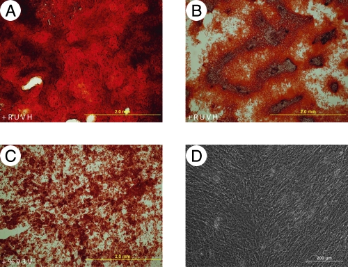
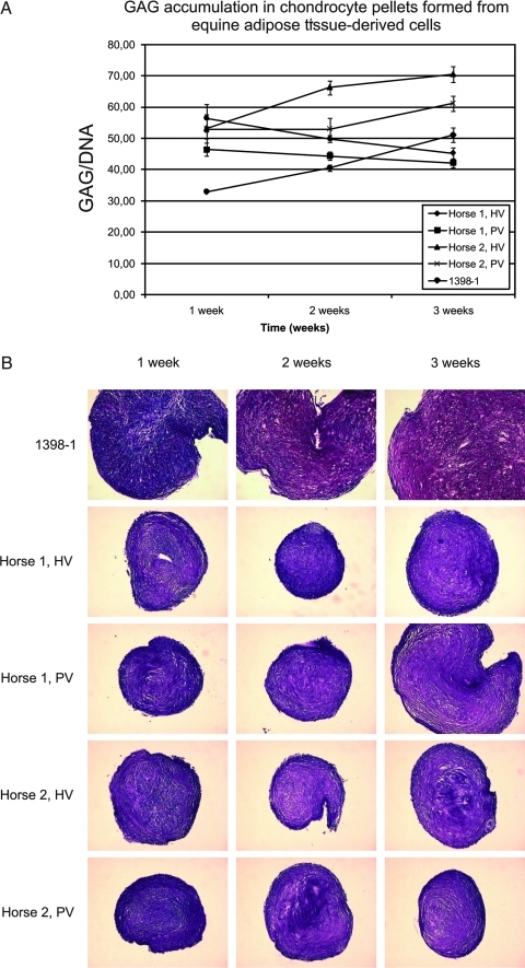
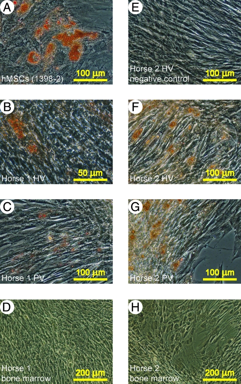
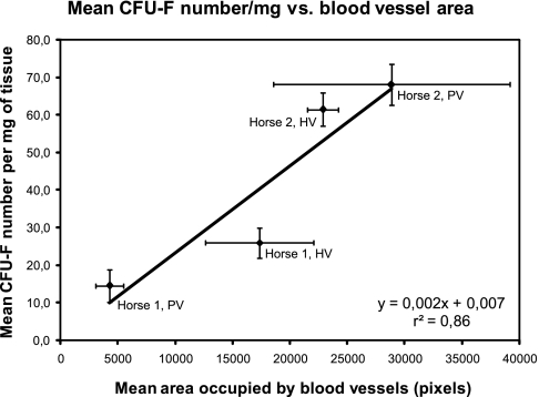
Mesenchymal stem cells (MSCs) are multipotent cells that have the capacity to develop into different mature mesenchymal cell types. They were originally isolated from bone marrow, but MSC-like cells have also been isolated from other tissues. The common feature of all of these tissues is that they all house blood vessels. It is, thus, possible that MSCs are associated with perivascular locations. The objective of this work was to test the hypothesis that MSCs are associated with blood vessels by verifying if MSC frequency positively correlates with blood vessel density. To this end, samples from highly and poorly vascularized adipose tissue sites of two equine donors were collected and processed for histology and cell isolation. MSC frequency in these samples was estimated by means of CFU-F assays, which were performed under MSC conditions. Culture-adherent cells from equine adipose tissue and bone marrow were culture expanded, tested for differentiation into mesenchymal cell types in vitro, and implanted in vivo in porous ceramic vehicles to assess their osteogenic capacity, using human MSCs and brain pericytes as controls. The differentiation assays showed a difference between adipose tissue-derived cells as compared to equine bone marrow MSCs. While differences in CFU-F frequencies between both donors were evident, the CFU-F numbers correlated directly with blood vessel densities (r(2) = 0.86). We consider these preliminary data as further evidence linking MSCs to blood vessels.
Figures
References
-
- Caplan A.I. Mesenchymal stem cells. J Orthop Res. 1991;9:641. - PubMed
-
- Caplan A.I. Dennis J.E. Mesenchymal stem cells as trophic mediators. J Cell Biochem. 2006;98:1076. - PubMed
-
- Haynesworth S.E. Goshima J. Goldberg V.M. Caplan A.I. Characterization of cells with osteogenic potential from human marrow. Bone. 1992;13:81. - PubMed
-
- Pittenger M.F. Mackay A.M. Beck S.C. Jaiswal R.K. Douglas R. Mosca J.D. Moorman M.A. Simonetti D.W. Craig S. Marshak D.R. Multilineage potential of adult human mesenchymal stem cells. Science. 1999;284:143. - PubMed
-
- Mosca J.D. Hendricks J.K. Buyaner D. Davis-Sproul J. Chuang L.C. Majumdar M.K. Chopra R. Barry F. Murphy M. Thiede M.A. Junker U. Rigg R.J. Forestell S.P. Bohnlein E. Storb R. Sandmaier B.M. Mesenchymal stem cells as vehicles for gene delivery. Clin Orthop Relat Res. 2000;379:S71. - PubMed
Publication types
MeSH terms
Substances
LinkOut - more resources
Full Text Sources
Other Literature Sources
