Review
. 2009 Jan;1788(1):64-71.
doi: 10.1016/j.bbamem.2008.09.003. Epub 2008 Sep 20.

Domain coupling in asymmetric lipid bilayers

Affiliations
Review

Domain coupling in asymmetric lipid bilayers

Volker Kiessling et al. Biochim Biophys Acta. 2009 Jan.

Abstract

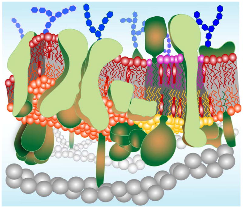
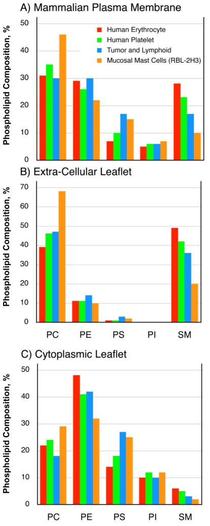
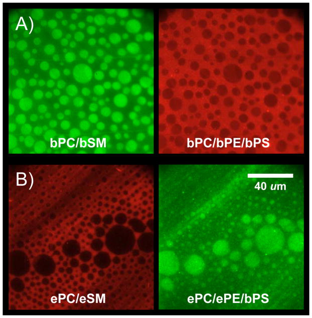
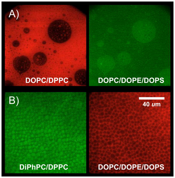
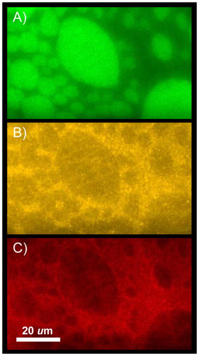
Biological membranes are heterogeneous assemblies of lipids, proteins, and cholesterol that are organized as asymmetric bimolecular leaflets of lipids with embedded proteins. Modulated by the concentration of cholesterol lipids and proteins may segregate into two or more liquid phases with different physical properties that can coexist in the same membrane. In this review, we summarize recent advances on how this situation can be recreated in a supported bilayer format and how this system has been used to demonstrate the induction of ordered lipid domains in lipid compositions that are typical for the inner leaflet by lipid compositions that are typical for the outer leaflet of mammalian plasma membranes. Proteins are shown to differentially target such induced inner leaflet domains.


Figures

Figure 1
Figure 1
A contemporary model of a biological membrane. Lipids are organized in a dynamic bimolecular film with asymmetry across the membrane and a lateral organization in cholesterol-rich and cholesterol-poor domains. The outer leaflet (purple and red) is enriched in sphingomyelin and the inner leaflet (orange and yellow) is enriched in phosphatidylethanolamine, phosphatidylserine, and phosphatidylinositol. Both leaflets contain similar amounts of phosphatidylcholine and cholesterol (lollipop-shaped red structures). Cholesterol-rich lipid domains (“rafts”) are shown in register in both leaflets as determined experimentally (see text). Transmembrane, monotopic (partially inserted), and lipid-anchored proteins (green) occupy much of the total available membrane area and are also clustered into functional complexes in many cases. Glycosylations on proteins and lipids of the outer leaflet are shown as blue hexagon-shaped structures. Gray beaded helical structures symbolize elements of the cytoskeleton, which interact with proteins on the cytoplasmic leaflet of the membrane.
Figure 2
Figure 2
Inner and outer leaflet lipid compositions of some mammalian plasma membranes. (A) Estimated total lipid headgroup compositions of the human erythrocyte (red, [36]), human platelet (green; [37]), tumor and lymphoid cell (blue; [38]), and mucosal mast cell (orange; [39]) plasma membranes. (B) Estimated lipid headgroup compositions of the extra-cellular leaflets of mammalian plasma membranes based on the works of refs. [36, 37]. (C) Estimated lipid headgroup compositions of the cytoplasmic leaflets of mammalian plasma membranes based on the works of refs. [36, 37].
Figure 3
Figure 3
Outer-to-inner leaflet coupling in asymmetric lipid raft domains formed from natural lipid mixtures. (A) Brain lipid system. The first leaflet of the supported bilayer (green) is composed of brainPC:brainSM:cholesterol (2:2:1) and labeled with lo-phase partitioning NBD-DPPE. The second leaflet (red) is composed of brainPC:brainPE:brainPS:cholesterol (1:1:1:0.75) and labeled with ld-phase partitioning rhodamine-DPPE. (B) Egg lipid system. The first leaflet of the supported bilayer (red) is composed of eggPC:eggSM:cholesterol (2:2:1) and labeled with ld-phase partitioning rhodamine-DPPE. The second leaflet (green) is composed of eggPC:eggPE:eggPS:cholesterol (1:1:1:0.75) and labeled with lo-phase partitioning NBD-DPPE. Adapted from ref. [33].
Figure 4
Figure 4
Outer-to-inner leaflet coupling in asymmetric lipid raft domains formed from synthetic lipid mixtures. (A) Unsaturated PC system. The first leaflet of the supported bilayer (red) is composed of DOPC:DPPC:cholesterol (2:2:1) and labeled with ld-phase partitioning rhodamine-DPPE. The second leaflet (green) is composed of DOPC:DOPE:DOPS:cholesterol (1:1:1:0.75) and labeled with lo-phase partitioning NBD-DPPE. (B) Saturated PC system. The first leaflet of the supported bilayer (green) is composed of DiPhPC:DPPC:cholesterol (2:2:1) and labeled with lo-phase partitioning NBD-DPPE. The second leaflet (red) is composed of DOPC:DOPE:DOPS:cholesterol (1:1:1:0.75) and labeled with ld-phase partitioning rhodamine-DPPE. Adapted from ref. [33].
Figure 5
Figure 5
Targeting of synaptotagmin C2A domains to induced inner leaflet lipid rafts in an asymmetric lipid system. The first leaflet of the supported bilayer (green) is composed of brainPC:brainSM:cholesterol (1:1:1) and labeled with lo-phase partitioning NBD-DPPE. The second leaflet (yellow) is composed of brainPC:brainPE:brainPS:cholesterol (1:1:1:1.33) and labeled with ld-phase partitioning rhodamine-DPPE. (C) 0.2 μM Alexa-647-labeled synaptotagmin I C2A (red) bound to asymmetric bilayer in the presence of 1mM Ca2+.

References

    1. Singer SJ, Nicolson GL. The fluid mosaic model of the structure of cell membranes. Science. 1972;175:720–731. - PubMed
    1. Engelman DM. Membranes are more mosaic than fluid. Nature. 2005;438:578–580. - PubMed
    1. Daleke DL. Regulation of transbilayer plasma membrane phospholipid asymmetry. J Lipid Res. 2003;44:233–242. - PubMed
    1. Ferguson-Miller S, Hochman J, Schindler M. Aggregation and diffusion in the mitochondrial electron-transfer chain: role in electron flow and energy transfer. Biochem Soc Trans. 1986;14:822–824. - PubMed
    1. McConnell HM, Vrljic M. Liquid-liquid immiscibility in membranes. Annu Rev Biophys Biomol Struct. 2003;32:469–492. - PubMed

Publication types

LinkOut - more resources