

Inner ear drug delivery for auditory applications
- PMID: 18848590
- PMCID: PMC2657604
- DOI: 10.1016/j.addr.2008.08.001
Inner ear drug delivery for auditory applications
Abstract
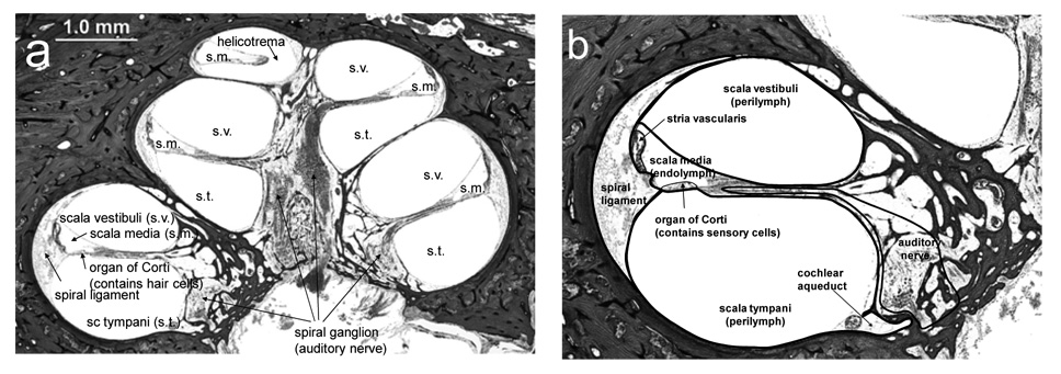
Many inner ear disorders cannot be adequately treated by systemic drug delivery. A blood-cochlear barrier exists, similar physiologically to the blood-brain barrier, which limits the concentration and size of molecules able to leave the circulation and gain access to the cells of the inner ear. However, research in novel therapeutics and delivery systems has led to significant progress in the development of local methods of drug delivery to the inner ear. Intratympanic approaches, which deliver therapeutics to the middle ear, rely on permeation through tissue for access to the structures of the inner ear, whereas intracochlear methods are able to directly insert drugs into the inner ear. Innovative drug delivery systems to treat various inner ear ailments such as ototoxicity, sudden sensorineural hearing loss, autoimmune inner ear disease, and for preserving neurons and regenerating sensory cells are being explored.
Figures
References
-
- Juhn S. Barrier systems in the inner ear. Acta Otolaryngol Suppl. 1988;458:79–83. - PubMed
-
- Juhn S, Rybak L. Labyrinthine barriers and cochlear homeostasis. Acta Otolaryngol. 1981;91:529–534. - PubMed
-
- McCabe BF. Autoimmune inner ear disease: Therapy. Am J Otol. 1989;10:196–197. - PubMed
-
- Moskowitz D, Lee KJ, Smith HW. Steroid use in idiopathic sudden sensorineural hearing loss. Laryngoscope. 1984;94:664–666. - PubMed
-
- Wilson WR, Byl FM, Laird N. The efficacy of steroids in the treatment of idiopathic sudden hearing loss. A double-blind clinical study. Arch Otolaryngol. 1980;106:772–776. - PubMed
Publication types
MeSH terms
Substances
Grants and funding
LinkOut - more resources
Full Text Sources
Other Literature Sources
