

Tissue engineering with meniscus cells derived from surgical debris
- PMID: 18848784
- PMCID: PMC2672194
- DOI: 10.1016/j.joca.2008.08.001
Tissue engineering with meniscus cells derived from surgical debris
Abstract
Objective: Injuries to the avascular regions of the meniscus fail to heal and so are treated by resection of the damaged tissue. This alleviates symptoms but fails to restore normal load transmission in the knee. Tissue engineering functional meniscus constructs for re-implantation may improve tissue repair. While numerous studies have developed scaffolds for meniscus repair, the most appropriate autologous cell source remains to be determined. In this study, we hypothesized that the debris generated from common meniscectomy procedures would possess cells with potential for forming replacement tissue. We also hypothesized that donor age and the disease status would influence the ability of derived cells to generate functional, fibrocartilaginous matrix.
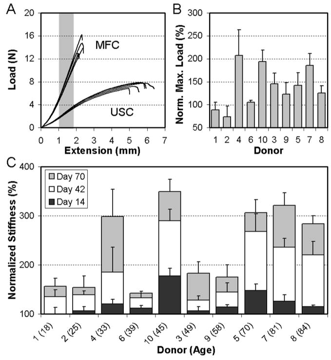
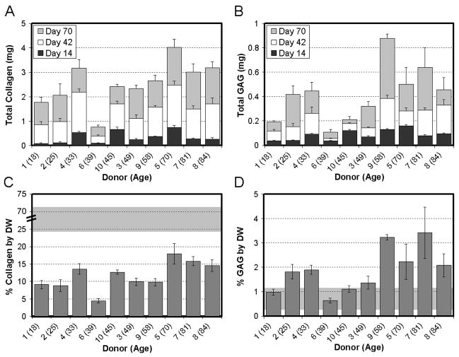
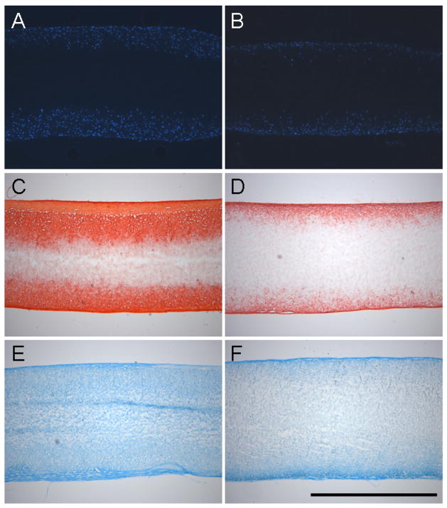
Methods: Meniscus derived cells (MDCs) were isolated from waste tissue of 10 human donors (seven partial meniscectomies and three total knee arthroplasties) ranging in age from 18 to 84 years. MDCs were expanded in monolayer culture through passage 2 and seeded onto fiber-aligned biodegradable nanofibrous scaffolds and cultured in a chemically defined media. Mechanical properties, biochemical content, and histological features were evaluated over 10 weeks of culture.
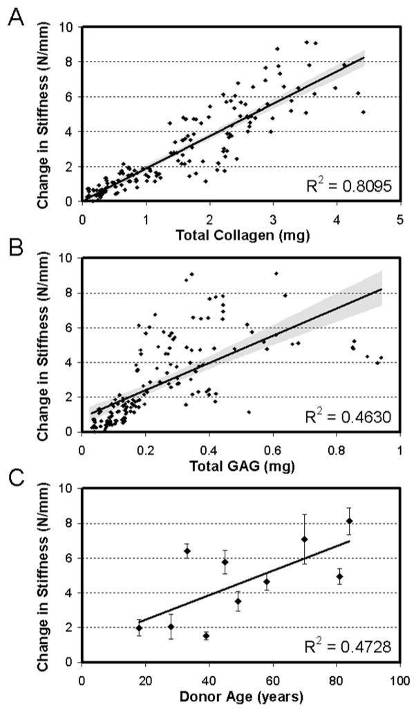
Results: Results demonstrated that cells from every donor contributed to increasing biochemical content and mechanical properties of engineered constructs. Significant variability was observed in outcome parameters (cell infiltration, proteoglycan and collagen content, and mechanical properties) amongst donors, but these variations did not correlate with patient age or disease condition. Strong correlations were observed between the amount of collagen deposition within the construct and the tensile properties achieved. In scaffolds seeded with particularly robust cells, construct tensile moduli approached maxima of approximately 40 MPa over the 10-week culture period.
Conclusions: This study demonstrates that cells derived from surgical debris are a potent cell source for engineered meniscus constructs. Results further show that robust growth is possible in MDCs from middle-aged and elderly patients, highlighting the potential for therapeutic intervention using autologous cells.
Figures
References
-
- Fithian DC, Kelly MA, Mow VC. Material properties and structure-function relationships in the menisci. Clin Orthop. 1990:19–31. - PubMed
-
- Kelly MA, Fithian DC, Chern KY, Mow VC. Structure and function of the meniscus: basic and clinical applications. In: Mow VC, Ratcliffe A, Woo SL-Y, editors. Biomechanics of Diarthrodial Joints. Vol. 1. New York: Springer-Verlag; 1990. pp. 191–211.
-
- Walker PS, Erkman MJ. The role of the menisci in force transmission across the knee. Clin Orthop Relat Res. 1975:184–92. - PubMed
-
- Ahmed AM. In: The load-bearing role of the knee meniscus. Knee meniscus: basic and clinical foundations. Mow VC, Arnoczky SP, Jackson DW, editors. New York: Raven Press, Ltd.; 1992. pp. 59–73.
Publication types
MeSH terms
Substances
Grants and funding
LinkOut - more resources
Full Text Sources
Other Literature Sources
